 History
A 17-year-old white male presented to the office with complaints of foreign body sensation, mild pain, tearing and redness of the right eye that had persisted for the last month. The patient had ulcerations on his lips as well as inside his mouth. He had no history of ocular trauma, disease or surgery, and reported no allergies or current medications.
Diagnostic Data
His best-corrected visual acuity through +0.50D spectacles was 20/100 O.D. at distance and near, which improved to 20/80 on pinhole testing. The vision in his left eye measured 20/20 at distance and near. His external examination––including cover test, pupils and confrontational visual fields––was normal. Refraction uncovered negligible changes.
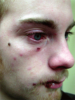
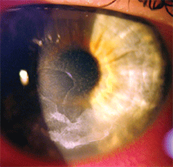
There was evidence of grade 2+ cell and flare O.D. His intraocular pressure measured 16mm Hg O.U. The dilated fundus examination revealed normal nerves and grounds with no peripheral pathology O.U. The immediately visible external signs are seen clearly in figure 1 and the pertinent biomicroscopic findings are illustrated in figure 2.
Your Diagnosis
How would you approach this case? Does this patient require any additional tests? What is your diagnosis? How would you manage this patient? What’s the likely prognosis?
Discussion
Additional testing included an evaluation of both corneas with fluorescein dye and superficial sensory testing as well as an analysis of the patient's facial muscles along the distribution of the right maxillary nerve. We uncovered reduced sensitivity of the right cornea compared to the left and reduced sensation of the right side of the face compared to the left. Muscular strength testing of both orbicularis muscles was also conducted along with an evaluation of the Bell’s reflex in each eye, which revealed some relative right-sided weakness.
The differentials to be considered in this case included herpes zoster ophthalmicus (HZO), keratoconjunctivitis, exposure keratopathy, neurotrophic keratitis, corneal abrasion and herpes simplex keratitis.


1,2. External photograph (top) and anterior segment image of our 17-year-old patient who presented with foreign body sensation, tearing and redness O.D.
The diagnosis in this case was HZO infection. Upon further questioning, it became clear that his medical history was complicated yet instrumental in understanding the underlying pathophysiologic mechanism. The patient explained that he experienced a right-sided earache three months earlier. After home remedies failed to alleviate the pain, the patient went to his general practitioner, who identified a substantial infection of his external and middle right ear (otitis external and media). The general practitioner immediately referred the patient to an otolaryngologist, who confirmed the diagnosis and discovered that early bacterial meningitis was manifesting. The patient was promptly hospitalized for IV antibiotic induction.
Unfortunately, the middle ear infection worsened and required surgical intervention. During the procedure, the right facial nerve was damaged, producing a postoperative motor deficit on the right side of the patient’s face. As a result of the meningitis, the opportunistic varicella organism induced an HZO infection on the right side of the patient’s face. After one month of treatment, his overall medical status improved and the patient was discharged from the hospital.
His primary care physician saw him after discharge and renewed his course of oral valacyclovir. Despite medical therapy, he had persistent vesiculobullous lesions on the right side of his face along with a red, irritated right eye. With no resolution following palliative topical therapy, he was referred to our office.
We found a large corneal epithelial defect infiltrated with calcium (band keratopathy) in the presence of a significant nongranulomatous iritis. We modified his topical regimen by adding Zymar (gatifloxacin, Allergan) q.i.d. to reduce the risk of concomitant infection within the area of the corneal break and also added scopolamine 0.25% b.i.d. for cycloplegic and analgesic benefit. Given the symptoms and severity of the injury, we believed that a bandage contact lens wasn't required.
The patient returned for follow up within 48 hours. The corneal defect was smaller and the eye was significantly less red. The anterior chamber reflected the healing process, exhibiting marked debris reduction. Also, his best-corrected visual acuity improved to 20/60 O.D.
With the corneal epithelium now progressing toward homeostasis, we decided to address the inflammatory aspects of the case. We added Lotemax (lotoprednol, Bausch + lomb) q.i.d. We then obtained a consult from a corneal specialist to rule out neurotrophic keratopathy and to evaluate the need for a tarsorrhaphy. The corneal specialist agreed with our diagnosis and treatment plan, and indicated that no additional procedures would be required.
Two months after the patient first presented, the corneal break and iritis had fully resolved and the vesicles were all but gone. Furthermore, his best-corrected visual acuity improved to 20/30 O.D.
The classic signs and symptoms of HZO produce characteristics that are virtually unmistakable. Vesicular lesions and a rash typically affect one dermatome of one side of the face. However, the entity may affect up to three dermatomes of the ipsilateral trigeminal nerve (cranial nerve V). The rash often starts as an erythematous area that increases in size along the affected dermatome(s). Soon after the rash appears, maculopapular lesions become evident and develop into vesicles or fluid-filled cysts. In 70% to 90% of patients, the rash is preceded by prodromal pain.1-7
It is also common for patients with herpes zoster to have a prodrome of fever, malaise, headache and dysesthesia for one to four days prior to the development of any visible cutaneous involvement. In fact, it may take 48 to 72 hours after the onset of pain for any sign of rash to manifest. Anecdotally, cases have been known to produce the painful prodrome over months without iritis, uveitis or vesicular breakout, making the chief complaint a mystery until the skin erupts. After the vesicles appear, they begin to collapse and crust for a period of seven to 10 days. This is part of the healing process and typically is followed by complete epithelialization. The lesions of herpes zoster generally resolve and heal completely within one to three weeks.1-6
The uncommon, but serious complication of meningoencephalitis has been documented.8,9 The exact incidence of herpes zoster meningoencephalitis is unknown; however, one study suggested that 5.5% of patients who initially presented with ophthalmic zoster also had additional neurological complications––one of which was meningitis.8 In another study of 26 patients with HZO, 50% had meningitis, 42% had encephalitis and 8% had acute disseminated encephalomyelitis.9 Interestingly, in this study, just 11 patients presented with the classic herpes zoster rash.9 HZO and its causative organism (varicella) are unquestionably associated with central nervous system disease, with meningitis being the most frequent clinical presentation.9 Even when the rash is not visible, the organism's presence must still be ruled out.
Thanks to Marc D. Myers, O.D., Jason Savatchka, O.D., and Brian Sirgany, O.D., for contributing this case.
1. Rogers RS, Tindall JP. Herpes zoster in the elderly. Postgrad Med. 1971 Dec;50(6):153-7.
2. Karbassi M, Raizman M, Schuman J. Herpes zoster ophthalmicus. Surv Ophthalmol. 1992 May-Jun;36(6):395-410.
3. Cobo M, Foulks GN, Liesegang T, et al. Observations on the natural history of herpes zoster ophthalmicus. Curr Eye Res. 1987 Jan;6(1):195-9.
4. Gungor IU, Ariturk N, Beden U, et al. Necrotizing scleritis due to varicella zoster infection: a case report. Ocul Immunol Inflamm. 2006 Oct;14(5):317-9.
5. De Freitas D, Martins EN, Adan C, et al. Herpes zoster ophthalmicus in otherwise healthy children. Am J Ophthalmol. 2006 Mar;142(3):393-9.
6. Gurwood AS, Savachka J. Herpes zoster ophthalmicus. Optom Today. 2001 Nov;41(11):34-8.
7. Watkinson S, Seewoodhary R. Managing the care of patients with herpes zoster ophthalmicus. Nurs Stand. 2011 Jun 1-7;25(39):35-40.
8. Srinivasan S, Ahn G, Anderson A. Meningoencephalitis-complicating herpes zoster ophthalmicus infection. J Hosp Med. 2009 Jul;4(6):E19-22.
9. Pahud BA, Glaser CA, Dekker CL, et al. Varicella zoster disease of the central nervous system: epidemiological, clinical, and laboratory features 10 years after the introduction of the varicella vaccine. J Infect Dis. 2011 Feb 1;203(3):316-23.

