 |
The Case
Dennis was diagnosed with a brain tumor in his fourth ventricle, which was removed in April 2012. After the removal, he had been seen at a local rehab hospital but suffered a fall and had meningitis. During his stay at the facility, he had his right eye patched by rehab staff because of double vision; prior to this, he simply closed his left eye all the time.
The patient was first seen in our clinic on June 19th, 2012, prior to beginning chemotherapy. He had no lens prescription and his unaided distance visual acuities were 20/30 OD, 20/40 OS and 20/30 OU. At near, unaided acuities were 20/60 OD, 20/40 OS and 20/30 OU. Cover testing at distance in his primary gaze showed a 35 prism diopter (PD) alternating esotropia with 10 PD of left hypertropia. At near, the horizontal component of the turn decreased to 30 PD. His eye movements were full, but he had trouble looking directly at the target and saw double in all fields of gaze. No mention of concomitancy vs. non-concomitancy was recorded. The retinoscopy exam yielded the following data:
• OD Plano -0.50 x 90 20/30.
• OS -0.25 -0.25 x 90 20/40.
We wondered why he wasn’t seeing better without lenses, given the retinoscopy findings. Often, the fixation skills of these patients have been so affected by brain problems that they achieve lower-than-normal visual acuities, but show improvement over time.
 |
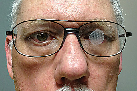
| Fig. 1. For patients who have lost CSBV due to TBI or ABI, sometimes the best outcome doesn’t revolve around getting it back. |
Attempting for CSBV
We measured the patient’s broken habitual Rx, which was:
• OD -1.00 -0.75 163 +2.00 add, 20/60 distance VA.
• OS -0.75 -0.75 14 +2.00 add, 20/40 distance VA.
First, we tried to help Dennis establish CSBV using a compensating prism. This approach allows the person to continue using their eyes in the turned position. We like to think of a prism used this way as shifting the images to where the eyes are. We often find that during periods of constant double vision, the patient is often quite agitated and cannot concentrate on looking or seeing. As a result, they fail to engage in the visual world around them. We first tried Fresnel prisms with 10 PD base up over the right eye and 35 PD base out over the left eye, over plano lenses in a distance-only frame. He returned on June 2nd and, based on his initial report that the prisms were indeed helping, the same prisms were put on +2.50 lenses for near. He reported that day seeing single vision with them.
Dennis returned on October 9th, after completing chemotherapy. He was still wearing his lenses, but reported blurred vision. Fresnel prisms do degrade the image seen through them—the higher the power, the worse the acuities. Since he had been closing his left eye prior to receiving prism, the higher amount was put over the left eye, degrading its acuity more than the right. The magnitude of the horizontal and vertical prism amounts was significant enough, and of high enough powers, that using one prism and rotating it to the right position, based on vector addition, was not a practical option. When it is practical, it allows us to rotate with the least amount of disruption.
We hoped that, as we helped him establish fusion, the binocular system would, on its own, become more robust and we could begin decreasing the prism amounts slowly over time. At the October 9th visit, Dennis was able to concentrate well enough that we obtained a better refraction, which gave him 20/25 acuity. Some myopia—approximately -1.00 OU—with a small amount of against-the-rule astigmatism was noted. The amount of prism needed had not changed, so we decided to begin vision therapy and prescribe two pairs of glasses, one for distance and one for near with the Fresnel prism.
A Change in Priority/Direction
Following 11 sessions of vision therapy, Dennis was reevaluated. We saw that he had a degree of non-concomitancy and, when he tracked from side to side, his two eyes were not moving smoothly together. The left eye was actually moving more smoothly than the right over most of the horizontal range. The left eye could not move far to the left without significant effort, and his right eye could not move far to the right. This led to him having to move his head to keep objects inside a narrow cone of vision.
Since he continued to complain about his clarity of sight, and it appeared that non-concomitancy was the underlying issue keeping him from progressing, we undertook a radical change: The prisms were removed and a spot patch was placed over his left eye.
He immediately loved how he saw. He remarked at the clarity of his vision—which resulted primarily from removing the prisms. The right eye was designated as the uncovered eye, because he still tended to close the left eye in the absence of prism. A spot patch, in this case a section of transpore tape, was placed on the front surface of the patient’s lens (Figure 1). This was done quickly and crudely as an experiment, and we checked him at his next scheduled vision therapy session. He loved the results of the transpore tape.
He could move his eyes and head freely now and, when he saw double, instead of shutting his left eye, he simply rotated slightly until the second image ducked under the spot patch. Over time, we moved the patch to the inside surface of the lens and, instead of transpore tape, we used Bangerter filters, which are more inconspicuous to the non-trained observer.

