 |
A patient may present to your office with reduced vision for any number of reasons, ranging from something as benign as uncorrected refractive error to serious pathology. Even in cases in which a combination of factors affect vision, the ability to distinguish between contributory ocular pathology and a refractive error is a must. Despite today’s high-tech tools—such as autorefractors or the potential acuity meter (PAM), to name a few—many ODs turn to a tried-and-true low-tech option: the pinhole occluder.
Optics Explained
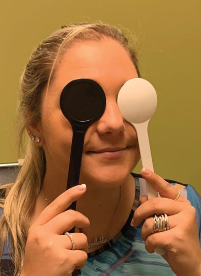
Pinhole occluders are inexpensive and readily available tools that consist of multiple, small apertures amidst a dark background. They are available for purchase or, in some cases, can even be fashioned from punching approximately 1mm to 2mm holes in a dark card.1
The pinhole occluder works along the same basis as pupil constriction in bright conditions causing an improvement in visual acuity. Through a smaller pupil, the effects of minor ocular irregularities—such as refractive error or paracentral cornea or lens opacities—are diminished.
The “pinhole effect” is an optical concept suggesting that the smaller the pupil size, the less defocus from spherical aberrations is present. When light passes through a small pinhole or pupil, all unfocused rays are blocked, leaving only focused light to land on the retina to form a clear image.1,2 In contrast, patients who have very large pupils, whether it is physiological or from pharmacological dilation, experience image “ghosting,” or a blurring halo around an otherwise sharp image.3
The size of the pinhole aperture or pupil size is an important consideration. While there is great benefit, as mentioned, to limiting marginal and unfocused rays from reaching the retina using a small aperture, a pinhole or pupil that is too small may cause diffraction and loss of resolution.1 Diffraction occurs when a light wave is fractured by the edge of the aperture, which limits the amount of detail available to form a clear retinal image.4 Additionally, a pinhole too small reduces retinal illumination. A clinical study evaluating pinhole size concluded that a diameter between 0.94mm to 1.75mm is most effective, with most instruments on the market containing approximately 1.2mm apertures.5
Pupil constriction is also involved in the near triad of accommodation, convergence and miosis. A balance of these factors is essential to increase depth of focus and aid in providing a clear near image. The effect of pupil constriction, or use of a pinhole, is such that the subsequent increase in depth of focus lessens the accommodative demand required.3
 |
| Due to its effects of limiting light scatter and subsequent blur, the pinhole can serve as a quick test to determine the need for further investigation of underlying ocular pathologies if the vision does not improve with pinhole. Click image to enlarge. |
Pinhole in Practice
The obvious benefit of using this optical principle in clinical practice is to discriminate between reduced visual acuity secondary to refractive error and the presence of pathology. When a patient’s visual acuity is not 20/20 despite the use of pinhole, further investigation to determine an underlying cause is warranted.
This is also a useful tool to check best-corrected visual acuity in patients for whom performing a refraction is unnecessary or difficult.6 In a practical setting, pinhole visual acuity may be used to document vision of a patient returning for a medical follow-up; routine refraction is not indicated, but sometimes the patient does not bring their up-to-date spectacle or contact lens correction to the appointment.
Another worthy application is in the context of measuring potential visual acuity post cataract extraction. Potential acuity pinhole (PAP) is a monocular test using a pinhole occluder to view a near target amidst bright illumination to predict visual status postoperatively.
The patient is first dilated so that they are able to search for a subjectively clearer area that may be less obstructed from lenticular opacities. Patients are then instructed to read the letters on a near card brightly lit with a transilluminator. The importance of using direct, bright lighting is to compensate for the reduced illumination from both the reduced viewing size and the cataract itself. The intensity of the light also helps to diminish the light scattering effect innate to cataracts.2,3 The best-corrected visual acuity measured using PAP correlates to the expected post cataract surgery visual outcome.
A similar test is the PAM test, which requires additional and more costly equipment for projection. In a study comparing the accuracy of PAP and PAM testing, both methods were similar in determining visual outcome post cataract surgery, though PAM provided a slightly more accurate correlation.3
Using pinhole visual acuity can offer the clinician a great deal of important information. It functions as a rapid tool to screen for best-corrected visual acuity without having to employ refractive techniques. It also suggests that if and when a pinhole fails to improve vision, the reduced vision is likely a result of pathology, whether a structural defect or functional change. Additionally, it is a readily available and cost-effective alternative to determine postoperative visual endpoints for patients undergoing cataract surgery.
1. Schiefer U, Michels R. Refractive errors: epidemiology, effects and treatment options. Dtsch Arztebl Int. 2016;113(41):693-702. 2. Melki S, Safar A, Martin J, et al. Potential acuity pinhole. Ophthalmology. 1999;106(7):1262-67. 3. Abdul N, van Bosch M, van Zyl A, et al. The effect of pinholes of different sizes on visual acuity under different refracting states and ambient lighting conditions. S Afr Optom. 2009;68(1):38-48. 4. Marcos S, Moreno E, Navarro R. The depth-of-field of the human eye from objective and subjective measurements. Vision Research. 1999;39:2039-49. 5. Borish IM. Borish’s Clinical Refraction. New York: Press books, 1988. 6. Sun JK, Aiello LP, Cavallerano JD, et al. Visual acuity testing using autorefraction or pinhole occluder as compared with a manual protocol refraction in individuals with diabetes. Ophthalmology. 2011;118(3):53-542. |

