The digestive and nervous systems frequently interact. Several disorders exist in which both gastrointestinal and ocular disease can occur. For instance, anterior uveitis may occur in association with inflammatory bowel disease. Both the gastrointestinal system and the eye may be involved as part of a systemic disease process. In addition, the gastrointestinal (GI) system may be adversely affected by the treatment of an unrelated eye disease.

Here, we describe the digestive system.
The GI Tract
The body’s digestive system breaks down food (i.e., carbohydrates, fats and proteins) into molecules small enough to be absorbed and transported by the circulatory system. These molecules are then distributed through cell membranes to provide the body’s cells with the energy required to properly function.
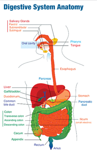
The gastrointestinal, or GI, tract (also known as the alimentary canal) includes the mouth, esophagus, stomach, small intestine, large intestine, rectum and anus. It’s a hollow, musculomembranous tube that is about 30 feet long in normal adults. The digestive system also includes glands and accessory organs (salivary glands, liver, gall bladder and pancreas).1 The liver, gall bladder and pancreas produce important secretions that drain into the small intestine.
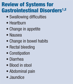
The gastrointestinal tract is a non-sterile system filled with bacteria and other flora. Common digestive disorders include gastroesophageal reflux disease (GERD), peptic ulcer and inflammatory bowel diseases.1,2 (See “Review of Systems for Gastrointestinal Disorders”)

Digestion
Digestion begins in the mouth. As food is chewed, saliva is secreted. Amylase, an enzyme, begins to break down starches. The form of amylase found in saliva is known as ptyalin.1
When food is swallowed, it enters the pharynx on its way through the esophagus. It is then transported to the stomach, where it mixes with gastric secretions, forming a semi-liquid known as chyme.1,2,3
The stomach consists on three parts: the fundus, body and antrum. The lining of the stomach secretes gastric juice that contains hydrochloric acid and the enzymes pepsin (which begins protein digestion), lipase (which speeds hydrolysis of emulsified fats) and in infants, rennin (which curdles milk so that pepsin can further break down proteins into polypeptides).1,2,3
Digestion mostly occurs in the stomach and in the duodenum of the small intestine. Peristalsis, a series of ring-like contraction waves beginning around the middle of the stomach, facilitates the mixing of chewed food with gastric juices. The stomach also produces an intrinsic factor responsible and necessary for the absorption of vitamin B12.4 The chyme remains mostly unabsorbed until it moves incrementally into the small intestine.
Absorption of nutrients occurs principally in the small intestine, a coiled tube consisting of the duodenum, jejunum and ileum. The stomach is continuous with the duodenum. The small intestine relies on many enzymes produced by the pancreas and by the intestinal lining itself to aid in digestion. Pancreatic enzymes include trypsin (digest protein to amino acids), lipase (digest fat to fatty acids and glycerol) and amylase (digests starches to sugars).3
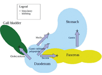
Action of Major Digestive Hormones

Hormones that help control digestion are produced and released by mucosal cells in the GI tract.
Intestinal enzymes include erepsin, lactase, maltase and sucrase.1 Bile, secreted by the liver and stored in the gall bladder, helps to neutralize stomach acid and to emulsify fats and fat-soluble vitamins, aiding the small intestine in the absorption process. The bile and main pancreatic ducts enter the posterior-medial duodenum.3
The large intestine consists of the cecum, appendix, colon, rectum and anal canal. Its function is to absorb water from the remaining indigestible food matter, and then to eliminate waste from the body.
The arterial supply to the digestive system is from the abdominal aorta. The portal vein collects blood from the abdominal part of the GI tract, pancreas, spleen and most of the gall bladder and carries it to the liver. (See “Digestive System Anatomy”)
The Role of Hormones

The gastrointestinal system and the eye may be involved as part of a systemic disease process.
The major hormones that control the functions of the digestive system are produced and released by cells in the mucosa of the stomach and small intestine. These hormones are released into the blood of the GI tract, travel back to the heart and through the arteries, and return to the digestive system where they stimulate production of juices and cause organ movement.
The main hormones that control digestion are gastrin, secretin and cholecystokinin (CCK).
• Gastrin. Gastrin causes the stomach to produce an acid for dissolving and digesting some foods. It is also necessary for normal cell growth in the lining of the stomach, small intestine, and colon.3
• Secretin. Secretin causes the pancreas to send out a digestive juice that is rich in bicarbonate. The bicarbonate helps neutralize the acidic stomach contents as they enter the small intestine. Secretin also stimulates the stomach to produce pepsin, an enzyme that digests protein, and stimulates the liver to produce bile.3
• CCK. CCK causes the pancreas to produce the enzymes of pancreatic juice, and causes the gallbladder to empty. It also promotes normal cell growth of the pancreas.3 (See “Action of Major Digestive Hormones”)
The Role of Nerves
Extrinsic and intrinsic nerves help control the action of the digestive system. Extrinsic nerves come to the digestive organs from the brain or the spinal cord. They release two chemicals, acetylcholine and adrenaline. Acetylcholine causes the muscle layer of the digestive organs to contract with more force and increase the “push” of food and juice through the GI tract. It also causes the stomach and pancreas to produce more digestive juice. Adrenaline, in contrast, relaxes the muscle of the stomach and intestine and decreases the flow of blood to these organs, slowing (or stopping) digestion.3
Intrinsic nerves make up a very dense network embedded in the walls of the esophagus, stomach, small intestine and colon. The intrinsic nerves are triggered to act when the walls of the hollow organs are stretched by food.3 They release many different substances that speed up or delay the movement of food and the production of juices by the digestive organs.
Together, the nerves, hormones, blood and organs of the digestive system conduct the complex tasks of digesting and absorbing nutrients from the foods and liquids that we consume.
Next: The Digestive System, Part II will highlight the ocular complications of gastrointestinal disease.
1. Chapter 4, Gastrointestinal Disorders. In: Professional Guide to Diseases, 9th ed. Philadelphia: Wolters Kluwer/Lippincott Williams & Wilkins; 2009: 233-98.
2. Khan A, Lightman S. The eye in gastrointestinal disease. Hosp Med. 2003 Sep;64(9):548-51.
3. Scott AS, Fong E. Body Structures and Functions, 10th ed. Clifton Park, NY: Thomson/Delmar Learning; 2004: 349-68.
4. Hoffman R, Benz EJ Jr, Shattil SJ. Hematology: Basic Principles and Practice, 3rd ed. New York: Churchill Livingstone; 2000: 446-84.

