 |
|
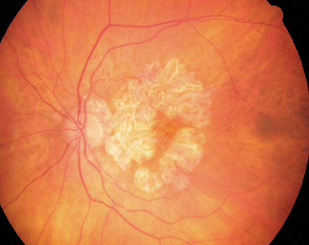
More precise tracking of geographic atrophy’s development can yield clues to its clinical course. Image courtesy of Wendy Harrison, OD, PhD. Click image to enlarge. |
Two prognostic tools that may help guide management decisions for age-related macular degeneration (AMD) patients with geographic atrophy (GA) include spectral-domain optical coherence tomography (SD-OCT) and fundus autofluorescence (FAF). A recent study examined several biomarkers for GA that can be measured through these imaging modalities to better inform treatment.
One hundred patients with GA underwent both SD-OCT and FAF imaging throughout a combined total of 1,062 visits, with follow-up lasting one to three years. Using a deep learning algorithm, the researchers quantitatively assessed hyperreflective foci (HRF) in SD-OCT volumes, which have been proven to represent activated retinal pigment epithelium cells migrating in an anterior direction within the retina. In FAF scans, they looked for specific FAF patterns that indicate faster development of new or enlargement of existing GA. FAF images were also observed for subretinal drusenoid deposits (SDD), GA lesion configuration and atrophy enlargement.
The researchers made the following conclusions:
- Eyes with higher HRF concentrations observed through SD-OCT showed a trend toward faster GA progression and revealed an impact on GA enlargement in interaction with FAF patterns.
- FAF patterns were significantly associated with GA progression.
- The diffuse-trickling FAF pattern presented significantly higher HRF concentrations, as well as higher mean sqrt GA growth, than any other pattern.
- SDD was associated with accelerated growth in existing GA. There was a faster growth rate of 0.1mm per 1mm atrophy growth per year in the presence of SDD.
- The condition of the fellow eye was not shown to have an effect on lesion enlargement.
These data represent some important biomarkers for monitoring the status and progression of GA, which is responsible for permanent vision loss in 20% of AMD patients who likely don’t receive timely testing and intervention.
“We found the presence of SDD and distinct FAF patterns to independently impact GA growth to the highest degree among a wide range of investigated biomarkers,” the study authors wrote. “Also, we demonstrated eyes with higher HRF concentrations to be significantly associated with GA enlargement in interaction with FAF patterns […] specifically, the diffuse-trickling pattern exhibited up to three-times higher concentrations of HRF than any other pattern.”
The authors concluded, “Identifying such disease markers using the combination of FAF and SD-OCT and precisely quantifying them using AI facilitates individualized patient management and enables more streamlined treatment trials, as well as deepens our understanding of the complex multifactorial nature of non-neovascular AMD.”
Bui PTA, Reiter GS, Fabianska M, et al. Fundus autofluorescence and optical coherence tomography biomarkers associated with the progression of geographic atrophy secondary to age-related macular degeneration. Royal College of Ophthalmologists. August 16, 2021. [Epub ahead of print]. |

