 |
Q: A 72-year-old white male presents with a history of sudden-onset horizontal diplopia for the past two days. I cringe when I see people with double vision. Where do I start?
A: “Whenever a patient over 50 presents with sudden-onset horizontal and binocular double vision, immediately think cranial nerve six (CN-VI) palsy secondary to a vascular etiology,” says Emily Williams, OD, a resident in ocular disease at Omni Eye Services of Atlanta in Georgia.
CN-VI palsy, also know as abducens palsy, is the most common extraocular nerve palsy seen, with an incidence of 11 out of 100,000 patients. While 30% of cases are idiopathic, vascular etiologies represent the most common cause in nearly 35% of cases. In non-traumatic, isolated CN-VI palsies, the most common vascular etiologies include hypertension (28%) and comorbid hypertension and diabetes (17%).1
“Taking a close look at the patient’s history is key to narrowing the differential,” says Dr. Williams. She adds that practioners should ask about and look for the following:
- Systemic conditions (e.g., hypertension, diabetes and thyroid imbalance).
- Recent illness or trauma.
- Symptoms of Graves’ disease and convergence insufficiency.2
 |
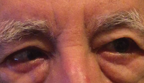
| When a patient over age 50 presents with sudden-onset horizontal and binocular double vision, immediately think CN-VI palsy secondary to a vascular etiology. Photo: Michael DelGiodice, OD. |
Dr. Williams says that when communicating with a CN-VI patient, observe for neurological symptoms indicative of prior vascular incidents like stroke. “And, always bear in mind the remote possibility of an etiology consisting in a benign or malignant growth.”
The key findings for determining CN-VI etiology typically manifest as the exam progresses. “Visual acuity, IOP, visual field results, pupils and slit lamp measurements should not be affected from an isolated CN-VI palsy,” says Dr. Williams. “Cover test will reveal a greater esotropia at distance than near and EOM testing will show the inability of the ipsilateral eye to abduct and will be accompanied by diplopia in that field of gaze.”
Dr. Williams also advises performing a dilated fundus examination to rule out specific vascular etiologies such as retinopathy from hypertension or diabetes.
 |
| This patient presented to the office with an abduction deficit in the right eye on right gaze. Photo: Michael Trottini, OD. |
In the absence of abnormal blood pressure, these patients do not need to be sent to the ER. “Educate the patient on the potential etiologies underlying their symptoms. If the patient has diabetes, it is worth mentioning that reoccurrence of the palsy is certainly possible.”
Dr. Williams advises telling patients it is essential to follow up with their PCP or internist to monitor uncontrolled systemic disease such as diabetes and hypertension as was seen in her patient. “This particular patient was sent back to his internist with my recommendation to evaluate for diabetes and to assess the adequacy of the type and dosage of his hypertension medication,” Dr. Williams says. The request was ignored and the patient sent for an MRI, which came back negative. His palsy resolved within nine weeks of the intital visit.
She says to inform your patient that if symptoms worsen, new symptoms begin, or if symptoms do not resolve within 12 weeks, head and orbital imaging will be needed. “The patient can return to your office six weeks from the onset of initial symptoms. A second follow up 12 weeks later should be scheduled to confirm resolution of the diplopia.”|
1. Goodwin D. Differential diagnosis and management of acquired sixth cranial nerve palsy. Optometry. 2006; 77(11):534-39. 2. Tasman W. Jaeger E: Abducens Palsies. Duanes: Clinical Ophthalmology. Volume 2. 12:3-9. |

