During our training and clinical careers––now spanning approximately 30 years––we have heard or read about a number of seemingly rare ocular events. In some cases, we’ve even taught them to our students and residents.
 |
Being honest, we learned about some of these events from our professors––but often have not stopped to think if we have actually seen them ourselves. This leads us to wonder if these occurrences are real or merely urban legends propagated through time.
This month we shine a light on some of these possible “ocular urban legends.”
Changing Parapapillary Atrophy?
We have long known that the optic disc goes through a number of characteristic changes in glaucoma. Focal rim damage, optic disc hemorrhages and loss of retinal nerve fiber layer (RNFL) all occur throughout glaucoma progression. We also know that parapapillary atrophy is associated with glaucoma––especially when occurring in the beta zone.1
Disc hemorrhages occur more frequently in eyes with parapapillary atrophy, and usually in the area of greatest atrophy width.2 We also know that eyes with zone beta parapapillary atrophy experience more significant and faster RNFL thinning, as well as faster glaucomatous progression, than those without this atrophy.3-5
The question arises: Does parapapillary atrophy change in glaucoma, and if so, is it a sign of disease progression? One large, population-based study in China showed that the five-year progression rate of zone beta parapapillary atrophy was 8.2% and was significantly correlated with higher IOP, greater myopic refractive error and increased central corneal thickness, and occurred more commonly in glaucomatous eyes.6

|

|
|
|
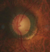
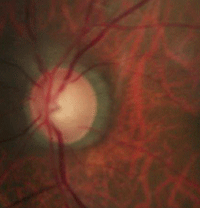
Our 63-year-old patient who exhibited parapapillary advancement over a five-year period in the left eye only (baseline examination left, five-year follow-up right).
|
||
Another research group noted that advancement of parapapillary atrophy occurred prior to optic disc or visual field change in some eyes with ocular hypertension, and may be an early finding in conversion to glaucoma.7
A separate team identified anatomic changes in parapapillary atrophy, but noted that detection was lower using standard ocular photos compared to alternation flicker technique (MatchedFlicker, EyeIc).8
One additional group reported that zone beta parapapillary atrophy does enlarge in glaucomatous eyes, but occurs very infrequently.9 They noted that this change occurs more often in patients with progressive glaucoma than in those with non-progressive glaucoma. They believed that due to low frequency, enlargement of zone beta may not be a very useful marker for glaucoma progression.9
Our thoughts? We’ll believe it when we see it.
The patient is a 63-year-old male treated for primary open-angle glaucoma since 2009. While his visual field and optic disc have not changed in either eye, his left eye shows a clear change in parapapillary atrophy. Because there is no consensus whether changing parapapillary atrophy is a sign of disease progression, we have not altered his therapy. We are following his corresponding visual field and optic disc photos (above) very carefully to see if they do change, before amplifying therapy.
Does Lens Removal Solve Pseudoexfoliation Syndrome?
Pseudoexfoliation presents as a fine, flaky material on the anterior lens capsule. Over time, this will coalesce into a characteristic “bulls-eye” pattern typically seen in pseudoexfoliation syndrome and glaucoma. Initially, intraocular pressure is unaffected in pseudoexfoliation syndrome; however, elevated IOP can develop in conjunction with glaucomatous cupping and visual field loss.
Pseudoexfoliation involves the production and accumulation of an abnormal fibrillar extracellular material within the anterior chamber of the eye. It appears that the material is comprised of abnormal basement membrane secreted by all structures within the anterior chamber, and is deposited on the anterior lens capsule, iris surface and trabecular meshwork.10,11 Due to accumulation of material at the pupillary margin, there is increased lenticular apposition with the iris and subsequent erosion of iris pigment as the pupil dilates and constricts.
The development of glaucoma typically occurs secondary to a buildup of pigment granules and exfoliative material within the trabecular meshwork. The primary cause of IOP elevation appears to be phagocytosis of accumulated pigment and material by the trabecular cells and Schlemm’s canal cells, with subsequent degenerative changes of Schlemm’s canal and the trabecular meshwork tissues.
The question arises: Does pseudoexfoliative material deposit on the intraocular lens or anterior capsule after cataract surgery? One research group noted this phenomenon to be a rare event, occurring in seven cases two to 20 years after lens removal.12 A second research team reported four cases in which pseudoexfoliative material deposited on the IOLs, with subsequent glaucoma development more than eight years after cataract surgery.13 Another group detailed a differing appearance in pseudoexfoliation deposition on the IOL surface, noting that the central homogenous zone is not present and that the material only deposits on the periphery of the IOL.14
Again, we’ll believe it when we see it.
The patient is a 75-year-old female who was treated for pseudoexfoliative glaucoma. She lives in the Bahamas and receives care there as well as here when she visits the US. She had visually significant cataracts at her last visit here 19 months ago. Prior to her return in 2014, she underwent cataract surgery in the Bahamas. At her most recent examination, we could clearly see pseudoexfoliative material on both the anterior lens capsule and the IOL. Thus, removing the lens did not solve pseudoexfoliation syndrome.
Does Exercise Cause IOP Spikes in PDS?
Patients with pigment dispersion syndrome (PDS) and pigmentary glaucoma demonstrate iris pigment liberation within the anterior chamber. The IOP rise in pigmentary glaucoma mostly occurs due to a breakdown of normal phagocytic activity in the endothelial cells and subsequent loss of normal trabecular architecture and function––not secondary to physical blockade of the meshwork.15
However, we have long heard tales of young PDS patients experiencing transiently blurred vision after exercising, presumably due to pigment liberation causing an IOP spike and resultant corneal edema. But does this really occur?
It appears that our understanding of this phenomenon comes from a single case reported in 1980.16 Another study group tried to recreate this phenomenon using a controlled exercise protocol involving 10 patients with PDS. While the researchers noted a minimal increase in both pigment liberation and IOP elevation 15 minutes after exercise, the pressures returned to baseline within 30 minutes.17 Two additional research teams noted that pigment liberation could be increased in patients with PDS after jogging and bicycling, but with no significant impact on IOP.18,19
One more time––we’ll believe it when we see it. And, for the record, we have never witnessed this phenomenon. Anecdotally, we have several academic colleagues who have tried to induce IOP elevation in their PDS patients to no avail. Thus, we don’t consider patients with PDS or pigmentary glaucoma to be precluded from exercise.
Indeed, there are phenomena that we have learned about but have never seen. While some may be rare events that do occur, others may just fall into the category of “urban legend.” Sometimes you don’t believe it until you see it.
1. Dai Y, Jonas JB, Huang H, et al. Microstructure of parapapillary atrophy: beta zone and gamma zone. Invest Ophthalmol Vis Sci. 2013 Mar;54(3):2013-8.
2. Radcliffe NM, Liebmann JM, Rozenbaum I, et al. Anatomic relationships between disc hemorrhage and parapapillary atrophy. Am J Ophthalmol. 2008 May;146(5):735-40.
3. Kim YW, Lee EJ. Microstructure of beta-zone parapapillary atrophy and rate of retinal nerve fiber layer thinning in primary open-angle glaucoma. Ophthalmology. 2014 Jul;121(7):1341-9.
4. Teng CC, De Moraes CG, Prata TS, et al. Beta-Zone parapapillary atrophy and the velocity of glaucoma progression. Ophthalmology. 2010 May;117(5):909-15.
5. Teng CC, De Moraes CG. The region of largest beta-zone parapapillary atrophy area predicts the location of most rapid visual field progression. Ophthalmology. 2011 Dec;118(12):2409-13.
6. Guo Y, Wang YX, Xu L, Jonas JB. Five-year follow-up of parapapillary atrophy: the Beijing Eye Study. PLoS One. 2012;7(5):e32005. Epub 2012 May 7.
7. Tezel G, Kolker AE, Wax MB. Parapapillary chorioretinal atrophy in patients with ocular hypertension. II. An evaluation of progressive changes. Arch Ophthalmol. 1997 Dec;115(12):1509-14.
8. VanderBeek BL, Smith SD, Radcliffe NM. Comparing the detection and agreement of parapapillary atrophy progression using digital optic disk photographs and alternation flicker. Graefes Arch Clin Exp Ophthalmol. 2010;248(9):1313-7.
9. Budde WM, Jonas JB. Enlargement of parapapillary atrophy in follow-up of chronic open-angle glaucoma. Am J Ophthalmol. 2004 Apr;137(4):646-54.
10. Amari F, Umihira J, Nohara M, et al. Electron microscopic immunohistochemistry of ocular and extraocular pseudoexfoliative material. Exp Eye Res 1997; 65:51-6.
11. Naumann GO. Pseudoexfoliation for the comprehensive ophthalmologist. Ophthalmology 1998;105:951-68.
12. da Rocha-Bastos RA, Silva SE, Prézia F, et al. Pseudoexfoliation material on posterior chamber intraocular lenses. Clin Ophthalmol. 2014;8:1475-8.
13. Park KA, Kee C. Pseudoexfoliative material on the IOL surface and development of glaucoma after cataract surgery in patients with pseudoexfoliation syndrome. J Cataract Refract Surg. 2007 Oct;33(10):1815-8.
14. Reiter C. Pseudoexfoliation syndrome: no central zone of pseudoexfoliation material in patients with pseudophakia––a clinical study. Klin Monbl Augenheilkd. 2012;229(3):241-5.
15. Richardson TM, Hutchinson BT, grant WM. The outflow tract in pigmentary glaucoma: A light and electron microcroscopy study. Arch Ophthalmol 1977; 95:1015-25.
16. Schenker HI, Luntz MH, Kels B, Podos SM. Exercise-induced increase of intraocular pressure in the pigmentary dispersion syndrome. Am J Ophthalmol. 1980 Apr;89(4):598-600.
17. Smith DL, Kao SF, Rabbani R, Musch DC. The effects of exercise on intraocular pressure in pigmentary glaucoma patients. Ophthalmic Surg. 1989;20(8):561-7.
18. Haynes WL, Johnson AT, Alward WL. Effects of jogging exercise on patients with the pigmentary dispersion syndrome and pigmentary glaucoma. Ophthalmology. 1992 Jul;99(7):1096-103.
19. Jensen PK, Nissen O, Kessing SV. Exercise and reversed pupillary block in pigmentary glaucoma. Am J Ophthalmol. 1995 Jan;120(1):110-2.

