History

A 77-year-old Asian female presented with a chief complaint of decreased vision in her left eye that had progressively worsened over the last year.
Her ocular history was remarkable for uncomplicated bilateral pseudophakia with yag capsulotomies approximately three years earlier.
She denied previous ocular injury or history of systemic disease. She was taking no medications and reported no allergies of any kind.
Diagnostic Data
Her best-corrected visual acuity was 20/50 O.D. and finger counting O.S. at distance and near. External examination revealed a grade II afferent pupillary defect O.S. There was evidence of a 70% loss in color vision, as well as decreased brightness sensation in her left eye.
Corneal examination found normal curvatures of 44.00D with clear mires in both eyes. Refraction yielded no improvement.

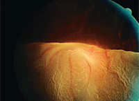
The left eye of our 77-year-old patient
who presented with a chief complaint of
decreased vision. What is your diagnosis?
The anterior segment exhibited well centered, securely placed intraocular lenses, with centered
capsulotomies and normal structures. The palpebral and bulbar conjunctiva were quiet and pink, without injection. There were no cells or flare in the anterior chamber.
Intraocular pressure measured 15mm Hg O.U. The pertinent posterior segment finding seen in the patient’s left eye is illustrated in the photograph.
Your Diagnosis
How would you approach this case? Does this patient require any additional tests? What is your diagnosis? How would you manage this patient? What’s the likely prognosis?
Discussion
Additional testing might include an amsler grid evaluation, Watske-Allen testing to evaluate macular integrity (which was positive), photodocumentation, B-scan ultrasonography and optical coherence tomography (OCT) testing.
The diagnosis in this case is a macular hole secondary to retinal stretching as a result of axial myopia. Her refractive error was corrected by the placement of IOLs; however, a resultant neurosensory retinal detachment developed that reduced her vision permanently. Because of a language barrier, it was difficult to instruct the patient to maintain steady forward fixation (even with an interpreter). This made it difficult obtaining clear diagnostic views during the physical examination. Most of the time, the macular area was seen just briefly as it flashed by or was observed through an opacified portion of the posterior capsule. Fortunately, photodocumentation permitted us to capture a clear image of the lesion, which helped us make an accurate diagnosis.
Macular holes may be induced secondary to idiopathic factors, chronic macular edema, partial macular tissue avulsion secondary to posterior vitreous detachment or tractional forces produced by epiretinal membrane formation.1-11 Macular holes may range from the incipient stages (where the chief presenting sign is a macular cyst with serous foveal detachment, resulting in only minor visual disturbances) to full thickness lesions with catastrophic endpoints.3,4
The current standard of treatment for early full-thickness macular holes is surgery, rather than observation.12-14 Surgical procedures have been shown to decrease the incidence of hole enlargement and can result in visual acuity improvement.12-14
The literature recognizes that some macular holes hold a poor prognosis for visual recovery.15 Old macular holes (generally more than 1.5 years), large holes (greater than 400µm) and holes that present secondary to another retinal pathology typically make significant visual function restoration more complicated.15
Classic complications following macular hole surgery include cataract formation, retinal pigment epithelium alterations, increased intraocular pressure, additional retinal breaks/detachment, macular hole enlargement, hole reopening, vascular occlusion, chronic CME, choroidal neovascularization, field loss and endophthalmitis.16,17
We referred the patient to a retina specialist for a three-port pars plana vitrectomy with internal limiting membrane peel and the insertion of expansile gas (perfluoropropane or sulfahexafluoride) with face-down positioning in an attempt to arrest the spread of the detachment and preserve peripheral vision in her left eye.1-3 While she refused to move forward with the procedure, her vision and retinal detachment stabilized with a final acuity of finger counting.
1. Kumagai K, Furukawa M, Ogino N, et al. Long-term outcomes of internal limiting membrane peeling with and without indocyanine green in macular hole surgery. Retina. 2006;26(6):613-7.
2. Krohn J. Duration of face-down positioning after macular hole surgery: a comparison between 1 week and 3 days. Acta Ophthalmol Scand. 2005;83(3):289-92.
3. Ang A, Snead DR, James S, et al. A rationale for membrane peeling in the repair of stage 4 macular holes. Eye (Lond). 2006;20(2):208-14.
4. Androudi S, Stangos A, Brazitikos PD. Lamellar macular holes: tomographic features and surgical outcome. Am J Ophthalmol. 2009;148(3):420-6.
5. Gass JDM. Reappraisal of biomicroscopic classification of stages of development of a macular hole. Am J Ophthalmol. 1995;119(9):752-59.
6. Kim JW, Freeman, WR, Azen SP, et al. Prospective randomized trial of vitrectomy or observation for stage 2 macular holes. Am J Ophthalmol. 1996;121(6):605-14.
7. Banker AS, Freeman WR, Kim JW, et al. Vision-threatening complications of surgery for full-thickness macular holes. Ophthalmology. 1997;104(9):1442-53.
8. Ezra E, Wells JA, Gray RH, et al. Incidence of idiopathic full-thickness macular holes in fellow eyes. A 5-year prospective natural history study. Ophthalmology 1998;105(2):353-59.
9. Nao-i N. Pearls in the management of macular holes. Seminars in Ophthalmology 1998;13(1):10-19.
10. Minihan M, Goggin M, Cleary PE. Surgical Management of macular holes: results using gas tamponade alone, or in combination with autologous platelet concentrat, or transforming growth factor β2. BMJ. 1997;81(12):1073-79.
11. Tsujikawa M, Ohji M, Fujikado T, et al. Differentiating full thickness macular holes from impending macular holes and macular pseudoholes. BMJ. 1997;81(2):117-22.
12. Carvounis PE, Kopel AC, Kuhl DP, et al. 25-gauge vitrectomy using sulfur hexafluoride and no prone positioning for repair of macular holes. Retina. 2008;28(9):1188-92.
13. Yagi F, Sato Y, Takagi S, et al. Idiopathic macular hole vitrectomy without postoperative face-down positioning. Jpn J Ophthalmol. 2009;53(3):215-8.
14. Farah ME, Maia M, Rodrigues EB. Dyes in ocular surgery: principles for use in chromovitrectomy. Am J Ophthalmol. 2009;148(3):332-40.
15. Susini A, Gastaud P. Macular holes that should not be operated. J Fr Ophtalmol. 2008;31(2):214-20.
16 Paques M, Massin PY, Santiago P, et al. Late reopening of successfully treated macular hoes. BMJ. 1997;81(8):658-62.
17. Hutton WL, Fuller DG, Snyder WB, et al. Visual field defects after macular hole surgery. A new finding. Ophthalmology. 1996;103(12):2152-59.

