 History
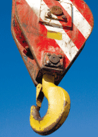
A 46-year-old black female presented for a soft contact lens evaluation. While taking her history, the patient complained of double vision for approximately one year since suffering blunt trauma at work. The patient is a heavy machinery operator at a port, and said that she was inadvertently struck in the area of her right temple by a cranes block and tackle.
She explained that the double vision was intermittent and occurred when she looked up and to the left, both with and without her spectacle correction. She did not complain of triplopia; however, she did report that the double vision remained when the right eye was covered in this field of gaze. The diplopia could not be alleviated with prisms and was described as diagonal (side-by-side component with an up-and-down component).
 The patient reported that her double vision was not present at near. She explained that she had visited countless doctors without satisfactory diagnosis or management. She disclosed records of her computed tomography scan, which showed a questionable hairline fracture of the right temporal orbit. She reported no additional medical history or previous ocular surgery.
Diagnostic Data
Her best-corrected visual acuity was 20/20 O.U. at distance and near through -3.75D-1.25Dx90 O.D. and -3.00D-0.75Dx90 O.S. correction. Keratometric readings demonstrated clear mires O.U., measuring 41.00D/41.75D in each eye. External examination found normal stereovision, color vision, confrontational visual fields and Amsler grid testing, with no afferent pupillary defect. There was no change in refraction.
Biomicroscopy uncovered normal external and internal structures. Her intraocular pressure measured 16mm Hg O.U. The dilated fundus examination was unremarkable, demonstrating normal nerves, maculae and peripheries.
Your Diagnosis
How would you approach this case? Does this patient require any additional tests? What is your diagnosis? How would you manage this patient? Whats the likely prognosis?
The Diagnosis and Discussion
In cases like this, additional evaluation may include a unilateral cover test in all positions of gaze, an alternating cover test in all positions of gaze, versions vs. ductions, and forced duction testing if an abnormality is observed. These tests will uncover any issues of entrapment or complications caused by damage to the third, fourth, sixth or seventh cranial nerves.
Topography and careful inspection of the cornea will uncover a related or unrelated refractive etiology, such as early keratoconus or budding corneal dystrophy. Gonioscopy will rule out angle damage and/or the potential for late traumatic glaucoma, in addition to lens dislocation, subluxation or opacity.
Exophthalmometry will reveal entrapment or proptosis of either eye. Also, carefully inspect the macula and vitreous to rule out retinal etiologies capable of producing unilateral optical distortion, such as old, unresolved commotio retinae or epiretinal membrane. Automated perimetry and additional neuroimaging are only indicated if a potential cause was defined.
The diagnosis in this case was corneal topographic changes O.S., presumed secondary to the old temporal head trauma.
The fourth cranial nerve is susceptible to trauma because it exits the brainstem ventrally and has the longest intracranial course.1 In our patient, however, the diplopia was still present when sensory fusion was eliminated. This prompted a topographical examination to gather data from the peripheral regions, since a keratometer only provides information on the central 2mm to 4mm of the corneal surface.2,3
The topographer mapped unusual irregularities in the left eye, which correlated with the visual field where the symptoms existed. We selected an appropriate rigid gas-permeable (RGP) trial contact lens and placed it on the eye to provide a uniform corneal surface.3 The RGP lens eliminated the symptoms and sharpened her vision.
We referred the patient to our corneal and contact lens service to rule out atypical keratoconus and/or other corneal disorders, and to finalize the RGP prescription. The patient did well with the rigid gas-permeable fitting, which should reduce the risk of fluctuation and/or additional changes.
1. Moster MM. Paresis of isolated and multiple cranial nerves and painful ophthalmoplegia. In: Yanoff M, Duker JS (eds.). Ophthalmology.
2. Mrochen M, Semchishen V. From scattering to wavefrontswhat"s in between? J Refract Surg 2003 Sep-Oct;19(5):S597-601.
3. Segal O, Barkana Y, Hourovitz D, et al. Scleral contact lenses may help where other modalities fail. Cornea 2003 May;22(4):308-10.

