She denied any recent illness, ocular infection or trauma. Her medical history was significant for hypertension and type 2 diabetes. She had moderate control of her blood sugar; her most recent fasting blood glucose level was 174mg/dL.

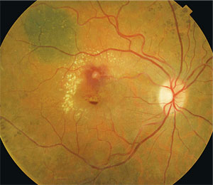
1. A fundus photograph of our patient’s right eye revealed several obvious changes. What do you see?  A 70-year-old white female was referred for evaluation following gradual, progressive vision loss in her right eye that began a month earlier. She reported that her vision had become exceptionally poor during the previous week.
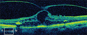
Fundus examination revealed obvious changes in her right eye (figure 1). We also performed an optical coherence tomography (OCT) scan (figure 2). Her left eye was normal, with no sign of background diabetic retinopathy or clinically significant macular edema (CSME).
Take the Retina Quiz
1. Based on the clinical images, what is the correct diagnosis?

2. An OCT scan of our patient’s right eye.
a. Vitreomacular traction (VMT).
b. Retinal arterial macroaneurysm (MA).
c. Diabetic retinopathy with CSME.
d. Macular telangiectasia.
2. What does the OCT reveal?
a. CSME.
b. Cystoid macular edema (CME).
c. VMT.
d. Stage II macular hole.
3. What is the traditional management strategy for this patient?
a. Observation.
b. Laser photocoagulation.
c. Pars plana vitrectomy with membrane peel.
d. Intravitreal Avastin (bevacizumab, Genentech).
4. What is the most likely cause of this patient’s condition?
a. Hypertension.
b. Diabetes.
c. Hyperlipidemia.
d. Both a and b.
5. What is the main cause of our patient’s reduced vision?
a. VMT.
b. CME.
c. Exudates.
d. Both a and b.
For answers, see below.
Discussion
Our patient presented with multiple retinal findings that contributed to her vision loss. On clinical examination, she had an MA located along the superior temporal arcade with surrounding exudation. Additionally, she had a mild epiretinal membrane that was less visible in the clinical photograph.
We expected that the OCT would show macular edema; however, we never imagined it would also reveal very obvious VMT. Finally, as an incidental finding, she had a choroidal nevus superior to the MA with overlying drusen.
MAs have been described in the literature since the late nineteenth century.1 They are acquired, usually round, dilatations of the retina’s large arterioles. Typically, they present unilaterally and occur within the first three bifurcations of the retinal arteries or at arteriovenous crossings in the superotemporal or inferotemporal vessels.2 Most patients with an MA will be asymptomatic. However, when the macula is involved, patients will experience painless vision loss.
MAs are commonly associated with exudates, edema, hemorrhages and neurosensory detachments––all of which may result in decreased visual acuity.3 While some associated exudates are clinically irrelevant, other exudates that are seen encircling MAs have the potential to cause significant vision loss. Additionally, MAs may present subretinally, intraretinally, preretinally or intravitreally. In any case, the visual prognosis depends on the amount of macular involvement and damage. Prolonged edema and hemorrhages can degenerate the retinal pigment epithelium and photoreceptors, causing permanent vision loss.4
MAs are most commonly seen in elderly individuals (aged 60 to 80 years), and are three times more likely to occur in females than males.5 Systemic risk factors include hypertension, atherosclerosis, hyperlipidemia, retinal arterial emboli, cerebrovascular disease and polycythemia.6 Hypertension is the most common systemic association, affecting approximately 75% of patients with MAs.7
Patients who present with MAs require a full investigation for concomitant systemic disease. Testing should include complete blood count (with differential and platelets), fasting blood sugar and glycosylated hemoglobin, blood pressure, fasting lipid profile, carotid auscultation or carotid duplex ultrasonography, and cardiac evaluation.8
Because MA is considered a “masquerade condition,” its clinical presentation can vary widely.5 Accordingly, differential diagnoses largely depend on the ocular signs observed on examination. If a vitreous hemorrhage is present, the differential diagnoses include proliferative diabetic retinopathy, retinal tear, branch retinal vein occlusion or hemorrhagic macular degeneration. If the hemorrhage is near or in the macula, consider macular degeneration, ocular histoplasmosis, high myopia, trauma, idiopathic polypoidal choroidal vasculopathy or choroidal neovascularization. Finally, if you observe retinal hemorrhages and exudates, the differential diagnoses include diabetic retinopathy, central or branch retinal vein occlusion, venous macroaneurysms, Coats’ disease, retinal telangiectasias, radiation retinopathy, cavernous hemangioma or retinal capillary angiomas.
Typically, fluorescein angiography (FA) can help confirm the diagnosis. On FA, MAs will be hyperfluorescent in the early phases and have a characteristic balloon appearance, and they will leak in the later phases.9
Current treatment options for MA include monitoring for spontaneous resolution, laser photocoagulation, laser photodisruption, vitrectomy and anti-VEGF treatments. The natural progression of most MAs includes spontaneous resolution without vision loss. A patient who presents with a non-leaking, asymptomatic MA should be monitored every four to six months. If the patient exhibits leakage with exudation and/or a hemorrhage that does not threaten the macula, monitor him or her every one to three months. If, however, you see a hemorrhage that threatens or involves the macula as well as any persistent edema, refer the patient for photocoagulation.
Additionally, any vitreous hemorrhage secondary to MA should be observed for three to four months. If spontaneous resolution does not occur, refer the patient for vitrectomy. In most cases, patients should regain all visual acuity unless the macula is affected by exudation or hemorrhage.
Even though laser photocoagulation is the most common treatment for MA, we administered an intravitreal Avastin injection to our patient at her initial visit. Our intention was to treat the macular edema associated with the MA as well as help release the VMT.
One month later, her central macular thickness decreased from 719µm to 496µm on OCT, and her visual acuity improved from 4/200 to 20/400 O.D. But, the vitreous did not detach itself. As a result, we scheduled her for a pars plana vitrectomy with membrane peel.
Thanks to Brianna Rhue, O.D., at Bascom Palmer Eye Institute in Miami, for contributing to this article.
Retina Quiz Answers: 1) b; 2) c; 3) b; 4) d; 5) d.
1. Doyne RW. Case of peculiar condition of the retina due possibly to the formation of small aneurysms and large extravasation of blood, which has become decolourised. Trans Ophthalmol Soc UK. 1896;16:94.
2. Humayun M, Lewis H, Flynn HW Jr, et al. Management of submacular hemorrhage associated with retinal arterial macroaneurysms. Am J Opthalmol. 1998 Sep;126(3):358-61.
3. Rabb MF, Gagliano DA, Teske MP. Retinal arterial macroaneurysms. Surv Ophthalmol. 1998 Sep-Oct;33(2):73-96.
4. Zhao P, Hyashi H, Oshima K, et al. Vitrectomy for macular hemorrhage associated with retinal arterial macroaneurysm. Ophthalmology. 2000 Mar;107(3):613-17.
5. Spalter HF. Retinal macroaneurysms: a new masquerade syndrome. Trans Am Ophthalmol Soc. 1982;80:113-30.
6. Lavin MJ, Marsh RJ, Peart S, et al. Retinal areterial macroaneurysms: a retrospective study of 40 patients. Br J Ophthalmol. 1987 Nov;71(11):817-25.
7. Sekuri C, Kayikcioglu M, Kayikcioglu O. Retinal artery macroaneurysm as initial presentation of hypertension. Int J Cardiol. 2004 Jan;93(1):87-8.
8. Kester E, Walker E. Retinal arterial macroaneurysm causing multilevel retinal hemorrhage. Optometry. 2009 Aug;80(8):425-30.
9. Moosavi R, Fong K, Chopdar A. Retinal artery macroaneurysms: clinical and fluorescein angiographic features in 34 patients. Eye (Lond). 2006 Sep;20(9):1011-20.

