 A 55-year-old white female presented for a routine eye examination. The patient suspected that she might need a new spectacle Rx. She had worn glasses for several years and had always enjoyed good vision; however, she reported a recent decline in her quality of vision. Her last eye exam was more than five years ago, and she did not recall any previous visual problems besides requiring glasses. Her medical history was unremarkable.
On examination, her best-corrected visual acuity measured 20/20 O.U. with a slight hyperopic correction. Extraocular motility testing was normal. Confrontation visual fields were full to careful finger counting O.U. Her pupils were equally round and reactive, with no afferent defect. Her anterior segments were normal. Her intraocular pressure measured 16mm Hg O.U.

|
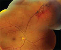
| 1. A fundus montage of our patient’s right eye. Note the peculiar lesion located along the superior nasal arcade. Look carefully in the posterior pole, where two other interesting findings can be seen.
|
Take the Retina Quiz
1. What do the peripheral retinal changes in the patient’s right eye represent?
a. Flame-shaped retinal hemorrhages.
b. Dilated telangiectatic retinal vessels.
c. Clusters of grape-like aneurysmal dilations.
d. Retinal neovascularizaton.
2. What is the correct diagnosis?
a. Congenital retinal telangiectasia (Coats’ disease).
b. Branch retinal vein occlusion (BRVO).
c. Cavernous hemangioma of the retina.
d. Choroidal hemangioma.
3. What do the changes in the posterior pole represent?
a. Retinal pigment epithelial detachments (PEDs).
b. Exudative retinal detachment.
c. Serous detachment of the retina.
d. Polypoidal choroidal vessels.
4. How should this patient be treated?
a. Observation.
b. Laser photocoagulation.
c. Intravitreal Avastin (bevacizumab, Genentech) injection.
d. Cryotherapy.
5. What is the likely evolution of the peripheral retinal changes?
a. No changes are likely.
b. Slow but continued resolution.
c. Leakage and exudation.
d. Continued growth.
For answers, see below.

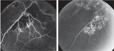
2, 3. The FA shows the mid (left) and late phases of the lesion in the patient’s right eye.
Discussion
Our patient has a cavernous hemangioma of the retina that is clearly visible along the superior nasal arcade. Cavernous hemangiomas of the retina are clusters of thin-walled, saccular aneurysms filled with dark venous blood that can resemble a cluster of grapes. These lesions are generally sessile, don’t change in appearance and don’t grow larger.1,2
Exudation is rare; however, small amounts of hemorrhage may develop, as was seen in our patient. In chronic cases, varying amounts of gray, fibrous tissue may develop on the surface. This appearance may increase over time.
Fluorescein angiography has shown that the sac-like tumors are relatively isolated from the usual retinal circulation. As a result, plasma-erythrocyte separation within the aneurysms is common (this finding has been referred to as a “pseudohypopyon”).1 In our patient, we see very slow and incomplete perfusion of the cavernous hemangioma without evidence of leakage. In the late phases of the angiogram, we observed some layering of the blood, which represents a separation of the plasma and erythrocytes.
Cavernous hemangiomas of the retina should not be confused with capillary hemangiomas. In capillary hemangiomas, the tumor develops as a part of the retinal circulation and has an afferent feeder vessel as well as an efferent drainage vessel. Capillary hemangiomas are subject to the stress of the normal retinal blood supply, whereas cavernous hemangiomas remain isolated because their blood supply is not part of the normal retinal vasculature.
Typically, cavernous hemangiomas of the retina are discovered as part of the routine eye exam. In most cases, the patient’s visual acuity is unaffected unless the tumor directly involves the macula. Nevertheless, visual disturbance occurs in approximately 10% of patients with cavernous hemangiomas.1 Patients may also have cavernous hemangiomas of the skin. And, in rare cases, the patient may experience central nervous system (CNS) involvement, particularly within the occipital cortex, brain stem or cerebellum, which may cause seizures or subarachnoid hemorrhages. Some investigators consider this complication to be part of a neuro-oculocutaneous syndrome.1,2
Nonetheless, because of the potential for CNS involvement, CT and/or MRI may be indicated—especially if there is a positive family history.1 Although no definitive pattern of inheritance has been established, autosomal dominance has been suggested.
In our patient, it is interesting to note the isolated blister-like retinal changes involving the posterior pole. There is one located temporally along the superior arcade and another located along the inferior arcade. These represent retinal pigment epithelial (RPE) detachments, which immediately light up on fluorescein angiography. There is no known association between cavernous hemangiomas of the retina and RPE detachments. So, it is more likely that these retinal changes represent a mild form of idiopathic central serous chorioretinopathy (ICSC) and are an incidental finding. Once again, because visual acuity is not affected, patients with such findings are usually observed without treatment.
As for our patient, we ordered an MRI to rule out CNV involvement. The work-up failed to show any CNS involvement. Her vision was unaffected by the cavernous hemangioma; she simply required an updated Rx. We will monitor her for significant changes every six months for the first year, and then yearly thereafter.
Retina Quiz Answers: 1) c; 2) c; 3) a; 4) a; 5) a.
1. Agarwal A, Aaberg TM Jr, Sternberg P Jr. Cavernous Hemangioma. In: Ryan SJ, Schachat AP (eds). Retina, Vol. I: Tumors, 4th ed. St. Louis: Mosby, 2006:609-13.
2. Gass JD, Stereoscopic Atlas of Macular Disease: Diagnosis and Treatment, 3rd ed. St. Louis: Mosby, 1987:634-8.

