 |
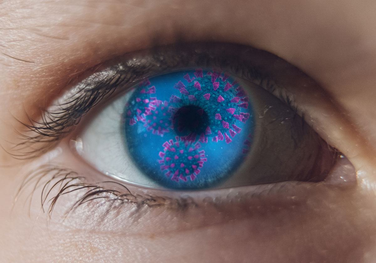
| Researchers observed patients who have recovered from COVID-19 still show vascular changes. |
SARS-CoV-2 has been detected in both the anterior and posterior chambers of the eye. Though there’s little evidence of posterior involvement, OCT-A has shown retinal microvascular changes in patients who recovered from COVID-19. To investigate the long-term sequelae of the virus, researchers recently assessed histopathological changes in the retina and choroid of affected donor eyes and observed severe subclinical anomalies.
The researchers examined seven COVID-19-positive eyes and six age-matched controls donor eyes ex vivo with macroscopic, scanning laser ophthalmoscopy and OCT imaging. They processed the macula and peripheral regions for Epon embedding and immunocytochemistry.
According to fundus analysis, the COVID-19 eyes showed more hemorrhagic spots and increased vitreous debris compared with healthy eyes. OCT revealed an increased trend in retinal thickness in COVID-19 eyes, but the difference wasn’t statistically significant. “Histology of the retina showed presence of hemorrhages and central cystoid degeneration in several donors,” the researchers noted. “Whole mount analysis of the retina labelled with markers showed changes in retinal microvasculature, increased inflammation and gliosis in the COVID-19 eyes compared with the controls. The choroidal vasculature displayed localized changes in density and signs of increased inflammation in the COVID-19 samples.”
“In this study, SARS-CoV-2 spike protein immunoreactivity showed different degrees of distinct and specific localization in round cells within the retina of all the COVID-19 eyes close to the optic nerve head,” the researchers said. “Additionally, our results show that both ACE2 (a cell surface receptor) receptor and TMPRSS2 (transmembrane protease serine 2) protein are present in the retina tissue of control donors. Thus, it remains a possibility that the mode of transmission for the virus could be through direct infection of the ocular tissue.”
“The anterior segments of our COVID-19 cohort’s eyes were previously analyzed,” they continued. “Of the 11 eyes recovered from seven COVID-19 donors, three conjunctival, one anterior corneal, five posterior corneal and three vitreous swabs tested positive for SARS-CoV-2 RNA. SARS-CoV-2 can cause anterior segment pathologies, including retinitis, optic neuritis, choroiditis with retinal detachment and retinal vasculitis. These studies further illustrate the importance of long-term assessments of ocular physiology of individuals who have recovered from COVID-19.”
Jidigam VK, Singh R, Batoki JC, et al. Histopathological assessments reveal retinal vascular changes, inflammation, and gliosis in patients with lethal COVID-19. Graefes Arch Clin Exp Ophthalmol. October 15, 2021. [Epub ahead of print]. |

