 History
A 35-year-old Asian male presented to the office following an emergency room visit for acute chemical exposure to both eyes. He was in considerable distress and reported altered vision. He had no known ocular history and reported no allergies.
Diagnostic Data
His best-corrected visual acuity measured 20/30 O.D. and 20/40 O.S. at distance and near. External examination was normal, with no evidence of afferent pupillary defect. The anterior segment examination uncovered grade III injection with coarse punctuate keratopathy O.U.
His intraocular pressure measured 19mm Hg O.U. The dilated fundus findings were normal.
Your Diagnosis
How would you approach this case? Does this patient require any additional tests? What is your diagnosis? How would you manage this patient? What’s the likely prognosis?
Discussion
Additional tests must include sodium fluorescein staining to assess the status of the cornea. Also, pH testing must be completed to determine the extent and type of toxicity. First aid mandates identification of the afflicting chemical as well as adherence to the suggested emergency exposure protocols for that substance. For example, some chemicals react with the oxygen found in water and should be brushed away. That is why proper identification is critical before initiating intervention. In most cases, the appropriate treatment is an immediate lavage with a neutral irrigating solution until the pH level is within normal limits. Also, it is optimal to use a Morgan Lens (MorTan Inc.) with a saline drip.
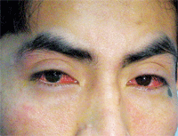
The diagnosis in this case is toxic conjunctivitis. Toxic conjunctivitis––sometimes referred to as toxic follicular conjunctivitis––results when the palpebral and bulbar conjunctivae have been acutely or chronically exposed to any number of foreign substances.1-4 Depending upon exposure, the process may occur unilaterally or bilaterally. Its clinical features include ocular itching; burning and tearing; injection of the bulbar and palpebral conjunctivae; chemosis; and inferior and/or superior eyelid follicle and papillae formation in the absence of preauricular lymphadenopathy.1-4 Keratitis formation is often present as well. Pannus formation may result in cases that are chronic, untreated or undertreated.4

External view of our 35-year-old patient who presented
following an acute chemical exposure.
There are many strata of ocular allergic reactions, so management is primarily aimed at reducing symptomatology.1-13 Perhaps the easiest and most effective treatment for allergic conjunctivitis is elimination or avoidance of the potentially offending allergen. Cold compresses, artificial tears and ointments can soothe and lubricate the ocular surface when applied on an as-needed basis.
Topical fourth-generation fluoroquinolones should be started q.i.d. to minimize the threat of infection. They can be withdrawn, without taper, once the cornea has been rehabilitated.
Topical combination medications, such as Patanol and Pataday (olopatadine 0.2%, Alcon), Zaditor (ketotifen fumarate, Novartis), Alaway (ketotifen fumarate, Bausch & Lomb), Optivar (azelastine hydrochloride, MedPointe) and Elestat (epinastine, Allergan), combine the effects of mast cell stabilization with antihistaminic properties to arrest symptoms immediately and increase resistance to additional episodes. Pataday provides the advantage of being a true once-a-day preparation.
Topical non-steroidal anti-inflammatory drugs (NSAIDs), such as Acular or Acuvail (ketorolac tromethamine, Allergan), Bromday (bromfenac, Ista Pharmacuticals), Nevanac (nepavenac, Alcon) and Voltaren (diclofenac, Novartis), may also offer relief in moderate cases of ocular allergy. Topical steroidal preparations, such as Lotemax (lotoprednol etabonate, Bausch & Lomb), FML Forte (fluorometholone 0.25%, Allergan), Pred Forte (prednisolone acetate, Allergan), Flarex (fluorometholone acetate, Alcon) and Inflamase Forte (prednisolone sodium phosphate, CIBA Vision), are typically reserved for non-remitting presentations.1-4 Durezol (difluprednate, Alcon) can be used in the most severe cases.
Our patient returned for his regularly scheduled, one-month follow-up examination and showed complete resolution. Fortunately, his vision returned to 20/20 O.U.
1. Fountain TR, Michon, JJ. Cornea/External Disease: Toxic Conjunctivitis. In: Varma R. Essentials of Eye Care: The Johns Hopkins Wilmer Handbook. Philadelphia: Lippincott-Raven; 1997:168-9.
2. Kairys DJ, Smith MB. General Principles of Antibacterial Agents. In: Onofrey BE (ed). Clinical Optometric Pharmacology and Therapeutics. Philadelphia: J.B. Lippincott Co.; 1994:1-25.
3. Groos EB. Complications of Contact Lenses. In: Duane TD, Tasman W, Jaeger EA (eds). Clinical Ophthalmology: Revised Edition for 1997. Philadelphia: Lippincott-Raven; 1997:1-21.
4. Dawson CR, Sheppard JD. Follicular Conjunctivitis. In: Duane TD, Tasman W, Jaeger EA (eds). Clinical Ophthalmology: Revised Edition for 1997. Clinical Ophthalmology: Revised Edition for 1997. Philadelphia: Lippincott-Raven; 1997:1-27.
5. Friedman NJ, Pineda R, Kaiser PK. Conjunctiva/Sclera: Conjunctivitis. In: Friedman NJ, Pineda R, Kaiser PK. The Massachusetts Eye and Ear Infirmary Illustrated Manual of Ophthalmology. Philadelphia: W.B. Saunders Co.; 1998:99-105.
6. Jackson WB. Differentiating conjunctivitis of diverse origins. Surv Ophthalmol. 1993 Jul-Aug;38 Suppl:91-104.
7. Onofrey BE. Soothing the symptoms of ocular allergy. Rev Optom. 1994;131(9): suppl.:6a -9a.
8. Jennings B. Mechanisms, diagnosis, and management of common ocular allergies. J Am Optom Assoc. 1990 Jun;61(6 Suppl):S32-41.
9. Onofrey BE. Principles of Immunology and The Immune Response. In: Onofrey BE (ed). Clinical Optometric Pharmacology and Therapeutics. Philadelphia: J.B. Lippincott Co.; 1994:1-13.
10. Kanski JJ. Disorders of The Conjunctiva. In: Kanski JJ. Clinical Ophthalmology: A Systemic Approach, 3rd ed. Boston: Butterworth-Heinemann; 1995:72-97.
11. Jackson WB. Differentiating conjunctivitis of diverse origins. Surv Ophthalmol. 1993 Jul-Aug;38 Suppl:91-104.
12. Cullom RD, Chang B. Conjunctiva/Sclera/External Disease: Allergic Conjunctivitis. In: Cullom RD, Chang B. The Wills Eye Manual: Office and Emergency Room Diagnosis and Treatment of Eye Disease. Philadelphia: J.B. Lippincott Co.; 1994:112.
13. Burden G, Bryant SA. Conjunctivitis. In: Burden G, Bryant SA. Laboratory and Radiologic Tests for Primary Eye Care. Boston: Butterworth-Heinemann; 1997:36-7.

