 |
In cases of acute posterior vitreous detachment (PVD), we need to be vigilant for any indication that a retinal problem may be present or imminent. Careful retinal exam is of utmost importance in determining the risk of any PVD-associated pathology, such as a tear, detachment or vitreomacular adhesion. While advanced diagnostic techniques require skill to master and are time-consuming to perform, there is also a quick, reliable way optometrists can identify a retinal tear or break: Shafer’s sign. Once you know what you’re looking for, you can’t miss it, and it might just make all the difference.
 |
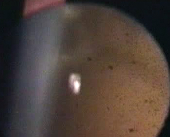
| Shafer’s sign, or “tobacco dust,” found in a slit lamp ophthalmoscopy test. Photo: Jeffrey Nyman, OD |
In the Wake of PVD
Acute PVDs occur in 63% of patients over age 69, and 18% of these cases will experience an associated retinal break.2 Typically, patients complain of flashes or floaters. The latter result from aggregation of collagen into visible fibers, blood in the vitreous cavity or the glial remnant (Weiss ring) following detachment around the optic disc. While the occurrence of flashes is less understood, they are likely due to vitreous traction, specifically the temporary separation of vitreous and retina induced by eye movements.3
While these common patient-reported symptoms are critical to the diagnostic process, a subsequent retinal exam is warranted in patients with acute PVD.4 Without taking immediate action, the persistent vitreoretinal traction that caused the initial tear can continue to lift the retina from underneath and cause a detachment.
To identify these breaks, eye care practitioners may choose to perform a retinal peripheral examination using three-mirror gonioscopy or binocular indirect ophthalmoscopy with scleral depression. However, these procedures require great clinical skill and practice, and that’s why we turn to Shafer’s sign for help.
Shafer’s Sign
Also called “tobacco dust,” Shafer’s sign refers to the presence of a collection of brown pigmented cells in the anterior vitreous following a PVD. First identified in 1965, the sign is best observed through slit lamp exam by sending a narrow, bright beam behind the posterior lens to focus on and illuminate the dark vitreous cavity.5 By finding and accurately identifying this collection of cells, the optometrist is often one step ahead of patient symptomology and can further secure their diagnosis of a retinal break.6 From here, it’s time to set out on the road to preventing a full detachment.
Table 1. Types of Cells Found in the Anterior Vitreous and their Clinical implications | ||
| Abnormal Vitreous Cells | Source | Clinical Indication |
| Brown (Shafer’s sign) cells | Pigment from RPE of retina | Retinal break |
| Red cells | Red blood cells from hemorrhage | Retinal break or proliferative retinal process |
| White cells | Inflammatory white blood cells | Vitritis, pars planitis |
Heed the Warning
While the origin of these pigment granules is unknown, they are thought to come from the shearing force of the break in the retinal pigment epithelium (RPE). During a PVD, as the vitreous forcefully tugs on the retina, the retinal break can occur. Next, the liquefied vitreous breaks down intercellular bonds in the RPE, causing brown pigment to be released into the vitreous cavity. The mark left by these free-floating cells is what we know today as Shafer’s sign.5,7 This theory has been supported by fluorescein angiography testing, revealing a break in the RPE in cases of anomalous PVD.7
Many studies suggest that Shafer’s sign is pathognomonic for a retinal break, assuming the patient has not previously undergone ocular surgery.1,2,8 If and when these pigment granules are observed in the vitreous, the likelihood of an associated retinal tear is 52 times higher than in cases where there is absence of these signs.2 Additionally, as 25% to 90% of retinal tears are expected to proceed to a retinal detachment, missing this warning sign could put your patient’s vision in grave danger.
Keep in mind that while the brown pigment granules in Shafer’s sign point to retinal breaks, red-pigmented cells in the vitreous will indicate vitreous hemorrhage. When these red cells are found following an acute PVD, there’s a 70% correlation with retinal tears. For this reason, both brown and red pigmented cells are causes for concern and further investigation.5 Instead, if white, or non-pigmented, cells are observed in the anterior vitreous, you are not dealing with a retinal break or tear. These white cells may indicate an inflammatory condition.7
Lastly, know that while the presence of Shafer’s sign confirms the presence of this associated retinal pathology until proven otherwise, the absence of Shafer’s sign in patients with acute PVD does not necessarily exclude the possibility of a retinal break or detachment.5 Additional testing should carry on as needed in these cases, whether or not their slit lamp exam shows the telltale tobacco dust.
Going Forward
While advanced technical procedures such as binocular indirect ophthalmoscopy with scleral depression may require years of practice, Shafer’s sign offers a quick, reliable method for identifying breaks and tears before they cause irreversible damage. As it can be easily identified through routine slit lamp ophthalmoscopy, all eye care practitioners possess the skill required to locate Shafer’s sign and heed its warning.
1. Bond-Taylor A, et al. Posterior vitreous detachment – prevalence of and risk factors for retinal tears. Clin Ophthalmol. 2017;11:1689-95. |

