 |
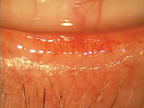
| Videokeratography could be an effective alternative to interferometers for assessing the lipid layer. Photo: Mile Brujic, OD. Click image to enlarge. |
Traditionally, interferometers have been used to assess lipid layer thickness (LLT) in patients with dry eye disease (DED) or meibomian gland dysfunction (MGD), as maintenance of the lipid layer is essential in promoting a stable tear film and healthy ocular surface. However, some interferometers rely on subjective diagnostic testing that could impact the accuracy of results. In a recent study, researchers examined a possible alternative method for assessing LLT involving the analysis of grey intensity values in videokeratography.
“During corneal topography measurement, the tear film acts as a mirror and reflects the projected placido disc ring pattern,” the authors of the study explained. “Placido disc rings appear lighter than the background. The healthy tear film surface forms a well-structured and reflected pattern with good intensity of reflection, while an altered tear film produces an irregular pattern with low reflectivity.” Therefore, an objective assessment of the lipid layer may be achieved by obtaining grey intensity values from the placido disc pattern.
The researchers examined the right eyes of 94 study volunteers aged 18 to 90 years old. Contact lens wearers were asked to refrain from wearing their lenses for a week leading up to examination. Using an Oculus Keratograph 5M to simultaneously record a video, the study team measured ocular surface parameters in participants, including the moment of the first breakup of the tear film (NIKBUT) and the average time of all breakups. The team then analyzed the video using an AI software called MatLab by MathWorks that was able to clearly isolate and highlight placido disc rings.
The analysis revealed stable pixel intensity of the placido disc in each subject throughout the measuring period that was unaffected by blinking. All recorded metrics showed acceptable repeatability and minimal variability, including total area under the pixel intensity curve, mean pixel intensity, standard deviation of pixel intensity, median pixel intensity and skewness.
“Moderate positive significant correlations were found between grey level intensities of the placido disc pattern, LLT and NIKBUT,” the researchers explained. “The correlations between new developed metrics, age, meibomian gland dropout, bulbar redness, tear meniscus height and OSDI might be a consequence of their correlation with LLT since LLT is also correlated with age, meibomian gland dropout and NIKBUT.” Despite new metrics being associated with LLT, it was not found to be statistically correlated with bulbar redness, tear meniscus height or OSDI.
Overall, analyzing grey level intensity values through videokeratography can help you assess the tear film in a quick, noninvasive way and objectively grade LLT. The metrics in the study were repeatable, and the researchers suggest this technique “could be easily included in a battery of tests to improve the detection and monitoring of DED and MGD in clinical practice.”
García-Marqués JV, Talens-Estarelles C, García-Lázaro S, et al. Validation of a new objective method to assess lipid layer thickness without the need of an interferometer. Graefes Arch Clin Exp Ophthalmol. August 9, 2021. [Epub ahead of print]. |


