 |
Q:
I don’t dilate my patients much anymore due to ultra-wide non-dilated photography. Is this the standard of care and am I at risk of missing something?
A:
“Although widefield photography is a useful screening tool, it has limitations,” says Robert Vandervort, OD, of Heartland Eye Consultants in Omaha, NE. He suggests that periodic dilated fundus examinations should always be performed in addition to any widefield screening.
Dr. Vandervort notes that, while the technology is improving, widefield imaging remains a screening tool that frequently does not allow proper examination of the peripheral fundus, especially in the superior and inferior portions of the retina. In addition, the lower magnification of widefield imaging reduces its value in assessing the subtle signs of vascular disease or early glaucomatous cupping.
On the other hand, the technology does provide the doctor with a stable image to review and, many times, picks up problems that might have been missed with a binocular indirect ophthalmoscope (BIO) or 90D fundus lens. However, it is not an outright replacement for a dilated fundus examination with binocular indirect ophthalmoscopy.
A Thorough Look
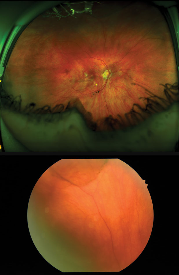
A patient was referred to Dr. Vandervort’s office after a routine eye examination for evaluation of chorioretinal atrophy in the macula. The patient was asymptomatic for any vision loss or any visual symptoms. Interestingly, while performing a dilated fundus examination of the right eye with BIO, he detected a superior retinal detachment (RD) with a pigment demarcation line in addition to the chorioretinal atrophy in the macula the referring doctor noted. Dr. Vandervort referred the patient to a local retina specialist for treatment with laser retinopexy.
Analyzing the widefield images sent to him, Dr. Vandervort noted that the inferior and superior retinal images provided limited views due to interference from the eyelashes and a narrow palpebral fissure width. This caused the retinal detachment to be obscured and go unnoticed. The macular chorioretinal scars were prominent and easily noted.
 |
| The standard fundus photo (below) was able to detect a superior retinal detachment that UWI (top) did not catch. |
The Appropriate Standard
Both patients and doctors have reasons to avoid dilation. Patients dislike the examination process and the resultant blurred vision and light sensitivity. Doctors would sometimes rather not interrupt a packed schedule by convincing patients of the value of dilation and the additional exam procedures needed to examine the fundus.While Dr. Vandervort agree sthat practitioners should be considerate of a patient’s wishes and comfort, he still recommends periodic dilation to avoid missing important and vision threatening findings. Widefield photos can be used in between dilated examinations, he adds.
“Just like any test we do, widefield imaging has value when appropriately balanced as part of the traditional examination techniques and tools we have available to us,” Dr. Vandervort explains. Overreliance on widefield imaging, and using it as a substitute for a thorough dilated fundus examination, can lead doctors into trouble.
Dilation is the still the standard of care, especially in higher risk patients. Dr. Vandervort advises that patients often have more than one condition —meaning that if they present with a red eye, don’t forget to examine the fundus. “When you need a thorough examination of the retina, there is no substitute for dilation by a capable and experienced clinician,” he adds.

