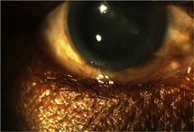
 A 60-year-old gentleman presents for evaluation, complaining of blurred vision in both eyes and chronic irritation of his right eye for the last six months. His medical history is significant for diabetes and hypertension, which are both treated pharmacologically with generic medications. His ocular history is reportedly unremarkable; he currently wears glasses for reading only. He claims to have tried several commercial eye drops from his local drugstore for the discomfort, but he says they afforded him little relief.
Examination reveals uncorrected visual acuity of 20/60 in both eyes, improving to 20/25 O.U. with a hyperopic refraction. Slit lamp examination shows age-consistent lenticular changes in both eyes. In the right eye, there is a slight inward turning of the lower lid with several large lashes in contact with the globe, creating an inferior corneal epitheliopathy that stains positively with sodium fluorescein.
Clearly, the patient’s issue is apparent, but how do we manage this condition effectively? This month, we’ll take a look at treating trichiasis.
Tricky Trichiasis

Trichiasis is defined as a misdirection of one or more eyelashes toward the globe. There may be a wide range of causes, clinical presentations and sequelae.
The primary etiology of trichiasis involves aging changes to the lids and lid margins, but scarring of the lid (from trauma, infection or inflammation) can also induce this condition. Trichiasis often results from entropion, in which the entire lid margin turns inward due to spastic or cicatricial changes.
In endemic regions of the world, trachoma—a chronic, infectious keratoconjunctivitis associated with the obligate intracellular bacterium Chlamydia trachomatis—is a major cause of trichiasis development.1 Other potential contributory conditions include Staphylococcal blepharitis, herpes zoster ophthalmicus, ocular cicatricial pemphigoid, Stevens-Johnson syndrome and chemical burns. Less commonly, misdirected lashes can be attributed to distichiasis, in which an extra row of follicles emanates from the meibomian gland orifices. Distichiasis is a metaplastic change that can be seen with inflammatory lid conditions.2
As the lashes chafe against the ocular surface, they cause irritation and inflammation. Corneal abrasion can ensue after prolonged or severe exposure, leading to further discomfort and the potential for microbial keratitis. Corneal scarring can result if this condition is not properly managed.2
Tools of the Trade
Mild cases of trichiasis can be handled with lubrication therapy, which serves to lessen irritation and protect the ocular surface from progressive damage. There are numerous ophthalmic lubricants on the market; however, when managing trichiasis, select a drop with medium to high viscosity to ensure adequate retention time. Some good options include Refresh Liquigel (Allergan), Systane Ultra (Alcon) and Genteal Gel (Novartis). Ointments can also be helpful in protecting the ocular surface, but typically induce too much blurring to be practical during daytime hours.
It’s also helpful to epilate misdirected lashes in the office, although this only provides temporary relief—eyelash follicles typically regenerate to full length within four to six weeks.2,3 Epilation is best accomplished at the slit lamp with jeweler’s or cilia forceps. Anesthesia isn’t necessary unless the patient is extremely apprehensive or the clinician is inexperienced.
Be sure to isolate the misdirected follicles and place the forceps at the base of the lashes, against the skin. With a firm grasp on the hair shaft just above the skin line, pull the lash from the follicle and make sure that the root emerges intact. Do not break the hair follicle along the shaft; this will leave a sharp, short “barb” that only induces more discomfort. A prophylactic antibiotic drop afterwards may help protect against any potential bacterial pathogens introduced by the procedure.
Recurrent or extensive trichiasis generally requires more invasive and definitive intervention. The hair follicle can be destroyed with electrolysis, which employs an electric current to prevent regrowth of the lash. Under local anesthesia, a fine probe is inserted approximately 2mm into the follicle, adjacent to the lash, and it coagulates the root of the hair shaft. The lash can then be removed harmlessly—and permanently—with forceps.
But, electrolysis is limited in that each lash must be treated individually, which makes for a time-consuming process. The therapy can also induce inflammation; treating a large number of lashes or using too much energy can potentially induce pain and even scarring. Perhaps the greatest limiting factor, however, is the tendency for electrolysis to fail in as many as 50% of cases.4
Laser photoablation has also been used successfully to correct trichiasis. Argon (blue-green, 514nm) laser was the first form of laser therapy to be used in this fashion; initial reports of such treatment were published more than 30 years ago.5 Since that time, refinements have included the use of infrared (810nm) diode lasers, Nd:YAG lasers (1064nm) and ruby lasers (694nm).6-8
Success rates for trichiasis management vary according to the method of photoablation used and the individual study, but overall, they range from 33% to 100%.8 Like electrolysis, laser treatment of trichiasis is a time-consuming process, particularly when low-energy lasers (e.g., argon) are used. Additionally, laser therapy is prone to similar complications including discomfort, inflammation, scarring and skin pigmentation changes.
Yet another treatment option for chronic trichiasis is cryotherapy, in which freon or nitrous oxide is used to destroy the errant follicles by freezing them. Cryotherapy has been used in the treatment of trichiasis for roughly the same duration as laser therapy.9 Advantages of this technique include low cost and short procedure duration.
Unfortunately, cryotherapy also has great capacity to cause “collateral damage” to nearby tissue. As many as 26% of patients demonstrate complications, including eyelid notching and scarring, pigmentary skin changes and destruction of normal eyelashes.10-11 Patients also tend to experience postoperative discomfort and inflammation.
In recent years, other approaches to trichiasis have been pioneered, including eyelash trephination, treatment with high-frequency radio waves, and a variety of surgical techniques.2,12-14 The success rates of these techniques are variable, but show great promise in the hands of experienced surgeons.
While there is no definitive therapy, know that numerous treatment options are available today beyond simple lubrication and epilation. Conditions like trichiasis are not typically seen as sight-threatening, but the long-term implications in terms of discomfort and potential ocular compromise cannot be ignored. Interventions ranging from artificial tears to laser and surgical treatments can be employed to reduce the morbidity of this condition, and it is the responsibility of the optometrist to facilitate such care for his or her patients.
1. Mabey DC, Solomon AW, Foster A. Trachoma. Lancet. 2003 Jul 19;362(9379):223-9.
2. McCracken MS, Kikkawa DO, Vasani SN. Treatment of trichiasis and distichiasis by eyelash trephination. Ophthal Plast Reconstr Surg. 2006 Sep-Oct;22(5):349-51.
3. Elder MJ, Bernauer W. Cryotherapy for trichiasis in ocular cicatricial pemphigoid. Br J Ophthalmol. 1994 Oct;78(10):769-71.
4. Chiou AG, Florakis GJ, Kazim M. management of conjunctival cicatrizing diseases and severe ocular surface dysfunction. Surv Ophthalmol. 1998 Jul-Aug;43(1):19-46.
5. Berry J. Recurrent trichiasis: treatment with laser photocoagulation. Ophthalmic Surg. 1979 Jul;10(7):36-8.
6. Başar E, Ozdemir H, Ozkan S, et al. Treatment of trichiasis with argon laser. Eur J Ophthalmol. 2000 Oct-Dec;10(4):273-5.
7. Pham RT, Biesman BS, Silkiss RZ. Treatment of trichiasis using an 810-nm diode laser: an efficacy study. Ophthal Plast Reconstr Surg. 2006 Nov-Dec;22(6):445-7.
8. Moore J, De Silva SR, O'Hare K, Humphry RC. Ruby laser for the treatment of trichiasis. Lasers Med Sci. 2009 Mar;24(2):137-9.
9. Hecht SD. Cryotherapy of trichiasis with use of the retinal cryoprobe. Ann Ophthalmol. 1977 Dec;9(12):1501-3.
10. Elder MJ, Bernauer W. Cryotherapy for trichiasis in ocular cicatricial pemphigoid. Br J Ophthalmol. 1994 Oct;78(10):769-71.
11. Zabriskie NA, Nordlund JJ, Nerad JA. Unusual skin depigmentation following eyelid cryosurgery. Ophthal Plast Reconstr Surg. 1996 Dec;12(4):296-8.
12. Kormann RB, Moreira H. [Treatment of trichiasis with high-frequency radio wave electrosurgery]. Arq Bras Oftalmol. 2007 Mar-Apr;70(2):276-80.
13. Rosner M, Bourla N, Rosen N. Eyelid splitting and extirpation of hair follicles using a radiosurgical technique for treatment of trichiasis. Ophthalmic Surg Lasers Imaging. 2004 Mar-Apr;35(2):116-22.
14. Moosavi AH, Mollan SP, Berry-Brincat A, et al. Simple surgery for severe trichiasis. Ophthal Plast Reconstr Surg. 2007 Jul-Aug;23(4):296-7.

