Dense asteroid hyalosis makes for difficult viewing of optic nerve details with normal funduscopy techniques in many cases. Evaluation of the optic nerve is critical for glaucoma patients. Here is where we can use some of the other tools in our diagnostic tool box to evaluate stability or progression of the disease.
Diagnostic Data
A 78-year-old Caucasian female presented in April for a glaucoma follow up. She had been a long-time patient who was initially observed as a glaucoma suspect, but converted to frank glaucoma about 12 years ago. At this visit, she was scheduled for threshold visual field studies and optic nerve imaging with multicolor imaging technology, as her dense asteroid hyalosis in her left eye rendered standard fundus photography useless.
In the past 12 years, she’s undergone slight changes to her medication regimen to facilitate stabilization. For the past four years, her glaucoma medications were pretty straightforward: generic latanoprost hs OU and 0.5% timolol Qam OU. Always a compliant patient, at this visit she asked an unusual question: “Why do I have to take the timolol since I’ve already had cataract surgery?” Interesting question from a long-standing glaucoma patient and one that got me thinking about medication confusion. I explained that the medication was for glaucoma, but I also asked her to tell me how and when she was administering her medications. She reported that upon awakening each morning, she would put a drop of the timolol into each eye, and then after about 15 minutes, she would put a drop of the latanoprost in each eye. Again, interesting evidence of dosing confusion.
 |
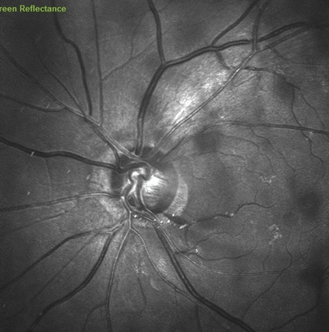
| Above, green laser image of the left optic nerve, demonstrating a disc hemorrhage at 4 o’clock and an adjacent wedge defect at 5 o’clock. Click image to enlarge. |
At this visit, entering visual acuities were 20/30- OD, OS and OU through hyperopic, astigmatic and presbyopic correction. These acuities were similar to her best corrected visual acuities obtained at the previous visit with minimal change in her refractive status. Pupils were ERRLA with no afferent pupillary defect noted.
Her anterior segments were essentially unremarkable, save for some mild endothelial guttatae in both eyes. Angles were open, and at the previous visit, gonioscopy demonstrated grade 4 open angles, with minimal trabecular pigmentation, and normal angle anatomy. Predilation intraocular pressures (IOP) were 12mm Hg OD and OS via Goldmann and central corneal thicknesses were previously measured to be 532µm OD and 518µm OS. Threshold visual fields were obtained. The patient was dilated prior to optic nerve imaging. Postdilation, adequate optic nerve images were obtained in her right eye, but not her left. When I looked at the blurred images of her left optic nerve, I remembered that she had dense asteroid. I proceeded to examine her and planned on obtaining her multicolor images.
Through dilated pupils, her posterior chamber IOLs were centered in the capsular bag, and the posterior capsules were open. She had long-standing bilateral PVDs. Her macular evaluations were stable with fine retinal pigment epithelium mottling in both eyes and minimal perifoveal drusen. Her retinal vascular status was stable with mild hypertensive and arteriosclerotic retinopathy OU.
Previously, her optic nerves were characterized by cupping of 0.55 x 0.70 OD and 0.55 x 0.65 OS. Examination of her left optic nerve was complicated by the asteroid, but with movement of the eye as well as the condensing lens at the slit lamp, I was able to obtain an adequate view of the left optic nerve. I noted a disc hemorrhage in the inferotemporal sector.
 |
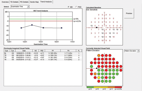
| The mean deviation trend analysis of the left visual field over 4 studies. Note MD is below statistical normal (~12db), given the density of her field defects, but appears to be stable over time. Click image to enlarge. |
Discussion
The presence of the disc hemorrhage in the left eye, being a new finding, called into question the overall stability of her optic nerve structure and function. In reviewing her medical record, I had noted two previous episodes of disc hemorrhages in previous years. But previous visual field studies and optic nerve imaging with both HRT 3 and OCT technologies demonstrated stable optic nerve appearances (at least in the past four years). The disc hemorrhage, along with the earlier discussion with the patient about her medication dosing, genuinely had me questioning the overall stability of her glaucoma, as the appropriate medication dosing would involve the timolol in the morning, and the latanoprost in the evening. Global trend analysis of the visual field in the left eye was stable.
At this point, I had one of my techs take the patient back to the OCT room and obtain the multi-color images, as well as an updated optic nerve glaucoma imaging protocol (which in my office includes three scans: a standard RNFL circle scan, an optic nerve radial scan, and a macular retinal scan with segmentation of all retinal layers), and asked the patient to be placed back in one of the examination rooms upon completion of the testing.
When I reviewed the new images and her visual fields, there was evidence that she had progressed. First, we obtained a decent multicolor image of her left optic nerve, clearly showing the disc hemorrhage. This will be helpful at the next visit to give me the sense of how quickly this hemorrhage resolves. OCT RNFL scanning demonstrated a significant focal reduction in the inferotemporal RNFL OS adjacent to the disc hemorrhage. This was also seen extending into the macular scan, in particular on the ganglion cell later segmentation.
An initial look at her visual field studies seemed to show no deterioration. Visual field interpretation is one of the most variable test results to interpret because so many factors are involved in obtaining accurate and reliable visual field studies. Quickly looking at an isolated field test can show how well preserved the visual field actually is, but it is only reflective of what is happening at that particular point in time. Where the real information lies is in the trend analysis that essentially looks at several field indices over time and statistically analyzes those data points, looking for an overall or a sectoral decline overtime.
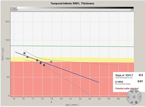
Being able to isolate individual sectors of the optic nerve in RNFL analysis (correlated to the Garway-Heath optic nerve sectors) is invaluable in dissecting out progressive structural damage in only one sector of the optic nerve, rather than a global deterioration of the RNFL.
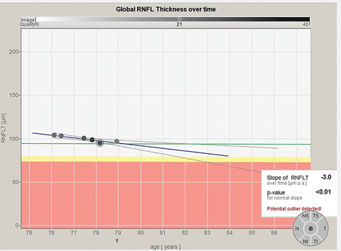
Remember, in this particular case, there is an isolated RNFL defect that has worsened. Is that isolated defect big enough to affect global RNFL indices? Or is it small enough where a subtle change in the RNFL would be statistically unnoticed when looking at global indices? As you can guess, the answer will vary, depending on the particular case, by how much damage has occurred structurally and where that damage has occurred. Similar to trend analyses in visual field interpretation, looking at RNFL data over time can give a clearer picture of how stable, or not, a patient remains.
 |
| Above, global optic nerve RNFL data over time. Note the slight decline in global RNFL thickness over time, implying slow progression. Below, inferotemporal Garway-Heath optic nerve sector RNFL data over time. Note the significant decline in the RNFL thickness in this one optic nerve sector, implying a rather significant progression. Click images to enlarge. |
 |
This case represents progression of the disease. In this instance, the progression is manifest as a focal RNFL defect, progressive RNFL and ganglion cell layer loss in the same area, with no significant change in her visual fields. The fact that her fields have remained relatively stable is certainly a good thing. But it is only a matter of time before the structural change will manifest as a field change.
Clearly, somewhere along the line, the patient began using both glaucoma medications in the morning, whereas they were initially prescribed to be dosed in the morning (timolol) and at bedtime (latanoprost). Many studies show IOP variability plays a role in progressive damage, and stabilizing IOP over a 24-hour period is important in preserving both structure and function. Did she begin using both medications in the morning two months ago, or two years ago? She really didn’t know, and neither do I. Did that play a role in the development of the RNFL defect? Possibly, and my guess is: probably. Will spreading the medication dosages out in the morning and the evening prevent further deterioration? Possibly, but only time will tell. Will she remain compliant, or will the gradual effects of aging make self-medication compliance more difficult? Possibly, but only time will tell.
Educate. Communicate. Maintain a healthy follow-up schedule. Monitor. Treat accordingly. Know that if things change in glaucoma, they only get worse, not better. Intervene early when needed. Monitor. And take solace in the fact that in most cases of glaucoma, time is on your side.

