 History
A 73-year-old black female presented for a routine follow-up examination six months after undergoing phacoemulsification with intraocular lens implantation in her right eye.
The patient explained that, while she was very happy with the outcome of the procedure, she noticed more glare at night and believed that her distance vision was not as good as it was immediately following the operation. She attributed this difficulty to the cataract in her fellow eye, and expressed a desire to have it removed.
The patient completed her postoperative dosing regimen, and denied use of any other medications. She reported no known allergies of any kind.
Diagnostic Data
Her best-uncorrected visual acuity was 20/40 OD, and her best-corrected visual acuity measured 20/25 OS. The external examination findings were normal, with no evidence of relative afferent pupillary defect.
Refraction revealed slight astigmatism in her right eye and mild myopia in her left eye, which was negligibly different than her habitual correction.
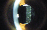
The anterior segment examination was normal. Intraocular pressure measured 17mm Hg OU. The dilated fundus examination was unremarkable and noncontributory. The pertinent slit-lamp findings are illustrated in the photograph.
Your Diagnosis
How would you approach this case? Does the patient require any additional tests? What is your diagnosis? How would you manage this patient? What is the likely prognosis?
Discussion
Additional testing could include pinhole visual acuity, laser interferometry to assess media-free macular capability and optical coherence tomography to insure macular integrity.

Slit-lamp view of our 73-year-old patient’s right eye. What do you notice?
The diagnosis in this case is posterior capsular opacification (PCO), or “Elschnig’s pearls.”
PCO is the most common long-term, postoperative cataract surgery complication.1,2 Advances in surgical techniques as well as intraocular lens (IOL) materials and designs have reduced the overall PCO rate; however, the condition still poses a significant problem.
PCO occurs when remnant lens epithelial cells are errantly left behind during surgery, permitting proliferation and migration. This leads to epithelial-mesenchymal transitioning, collagen deposition and new lens fiber generation on the remaining supportive structure of the posterior lens capsule.2 The process is influenced by cytokines, growth factors, extracellular matrix proteins and, according to some researchers, IOL geometry.1,2 The only effective treatment for PCO is Nd:YAG laser capsulotomy.2
Many studies have attempted to identify factors that influence the development of PCO. One study evaluated results from 26 prospective, randomized, controlled trials with a follow-up period of at least 12 months.1 In five out of seven grading categories, visual acuity was better in patients who were implanted with sharp-edged IOLs than in those who received round-edged IOLs. While the PCO score was markedly lower in patients with sharp-edged IOLs, there was no significant difference between one-piece and three-piece, open-loop IOLs.1 The authors concluded that, because of the substantial difference in PCO score, sharp-edged IOL optics should be employed more readily than round-edged IOL optics.1
Other researchers have hypothesized that lens material plays a role in the incidence of PCO.3 In one study that evaluated the outcomes of 90 eyes, 40% of eyes fit with hydrophilic IOLs exhibited PCO compared to just 8% fit with a hydrophobic IOL design.3 The authors concluded that hydrophilic acrylic IOLs appear to have a high incidence of PCO.3
Decreased vision following cataract surgery, as well as the formation of cystoid macular edema (CME) or Irvine-Gass syndrome, may result secondary to postsurgical inflammatory cytokine release.4 Optical coherence tomography (OCT) has the ability to reveal clinically relevant postoperative CME. While it should not be viewed as a replacement for fluorescein angiography, OCT is a major diagnostic advancement and adjunct.4
Eyes that develop clinically relevant CME exhibit substantial increases in macular thickness. A retinal thickness increase of at least 40% from baseline––documented via OCT imaging––may be the new, uniform standard for defining potentially treatable edema.4
New research conducted on rabbit eyes indicates that cyclosporine might be able to inhibit the occurrence of postoperative PCO.5 Cytotoxic medications also are under investigation for PCO prevention.
Our patient underwent a successful YAG capsulotomy in her right eye. Three days after treatment, her visual acuity improved to 20/20 OD.
1. Buehl W, Findl O. Effect of intraocular lens design on posterior capsule opacification. J Cataract Refract Surg. 2008 Nov;34(11):1976-85.
2. Awasthi N, Guo S, Wagner BJ. Posterior capsular opacification: a problem reduced but not yet eradicated. Arch Ophthalmol. 2009 Apr;127(4):555-62.
3. Saeed MU, Jafree AJ, Saeed MS, et al. Intraocular lens and capsule opacification with hydrophilic and hydrophobic acrylic materials. Semin Ophthalmol. 2012 Jan-Mar;27(1-2):15-8.
4. Kim SJ, Bressler NM. Optical coherence tomography and cataract surgery. Curr Opin Ophthalmol. 2009 Jan;20(1):46-51.
5. Pei C, Xu Y, Jiang JX, et al. Application of sustained delivery microsphere of cyclosporine A for preventing posterior capsular opacification in rabbits. Int J Ophthalmol. 2013;6(1):1-7.

