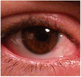
 Q: I have a patient who says he woke up two weeks ago with blurred vision in his left eye. His best-corrected visual acuity is 20/50 in that eye, 20/20 in the right eye. His exam is completely normal. What do I do next?
A: Good question, says optometrist Bill Woods, who has a very similar patient in his
Dr. Woods patient is a white 61-year-old male whose previous best-corrected acuity was 20/20 in both eyes. Now its 20/20 O.D., and 20/50 O.S.
He isnt hurting, Dr. Woods says. His eyes are not injected or irritated. Hes taking no medicines and has no headaches. His health is good. The only anomaly: a tiny internal chalazion on the left upper eyelid. Everything else in the exampupils, color, intraocular pressure, confrontation fields, Amsler gridwas negative.
First, Dr. Woods tried the pinhole test, which can point to subtle corneal changes such as early corneal map-dot-fingerprint dystrophy. This brought the patient down to 20/25 O.S. So, I tried to refract him and could not improve him at all.
Sometimes, patients just have a bad day and cant read 20/20. Theres nothing wrong with bringing them back and rechecking acuity. Just be sure to always make the appointment for them, whether its back to your office or to someone elses. So, Dr. Woods scheduled the patient for a second appointment.
But, when the patient came back two days later, there was still no improvement.
Dr. Woods reviewed the patients history. Again, the patient denied any injury or incident, and denied taking any medication that might cause blurriness. However, he did explain that the hordeolum had been larger two weeks prior, and he used hot compresses to bring it down. This was about when the blurriness began.
A subtle chalazion on the upper lid can cause unexplained vision loss.
Q: So, is the stye causing this patients visual acuity loss, or is it something more significant? Is further evaluation warranted?

A: Yes, further evaluation is necessary because, at worst, the vision loss could indicate a neurological problem.
But, the next logical step for this patient is topography to look for irregular astigmatism or perhaps early keratoconus.
If topography doesnt provide a clue, the next step is to place a diagnostic rigid lens on the eye with a drop of anesthetic. An over-refraction often results in 20/20 vision and solves the mysteryand it did so in Dr. Woods patient. A diagnostic lens brought the patient to 20/20, so we concluded that the stye was putting enough pressure on the cornea to create distortion and cause the patient some blurriness.
But, despite these efforts, what if the patient still had vision loss? In such a case, carefully examine the crystalline lens for occult milky nuclear sclerosis. Then, perform a careful dilated exam of the macula to rule out subtle serous retinopathy or cystoid macular edema.
If you suspect an optic nerve etiology, perform color vision testing and careful pupil testing.
Your final test is a visual field, and your best bet is a full (120- or 180-degree, not 30-degree) screening field for neurologic defects. If the field demonstrates any defect that respects the vertical midline, or you still dont have an explanation after running all these tests, send the patient for a neurologic consult with imaging to rule out an optic nerve or intracranial mass lesion.
Make the appointment for the patient. Then, send the visual fields and your findings along with another call to the neurologist explaining the case.
Fortunately for this patient, the only lump he had was a slowly, but surely, resolving one on his upper eyelid.

