Degenerative conjunctival lesions, such as pingueculae and pterygia, are frequently encountered in clinical practice. The diagnosis is easily made upon slit-lamp evaluation; corneal topo-graphy and external ocular photography can help document the progression. Treatment options include artificial tears, topical corticosteroids, topical cyclosporine, nutraceuticals, punctal occlusion and ultraviolet-blocking spectacles. In the event these prove ineffective, patients may need surgical intervention.

|

|
So, when is it time to refer for pterygium surgery? Whenever the condition “looks bad, feels bad or sees bad.” Indications include the presence of benign thickening of the outer conjunctiva that encroaches the cornea, loss of clarity within the visual axis, increasing corneal astigmatism, chronic irritation and inflammation, motility restrictions and cosmetic concerns. Any of these may warrant surgical referral.
In our cataract patients, the in-crease in corneal astigmatism may prompt referring ODs and surgeons to consider pterygium removal prior to cataract surgery, especially for patients with a spectacle-free refractive outcome in mind. The goal of cataract surgery is to improve the overall quality of vision. Removing the pterygium and waiting several months for the cornea to stabilize will allow for more accurate preoperative cataract surgery measurements. In our clinic, we typically wait four to eight weeks after pterygium removal before we schedule the cataract evaluation and surgery.
How it Goes
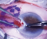
The procedure is performed in an outpatient setting under local (perhaps even topical) anesthesia and sedation; it typically takes less than a half hour. First, the borders of the pterygium are highlighted with a surgical marker or spot cautery to outline the area to be removed. The dissection then begins, at the corneal side of the pterygium. Using forceps, the head of the pterygium is lifted, dissected off the cornea in a lamellar fashion, and the remainder of the pterygium is removed with Wescott scissors to expose bare sclera.
 |
|
|
Click
here to see a video of pterygium removal.
|
Potential surgical techniques include bare sclera, conjunctival flap rotation and conjunctival autograft.
The bare-sclera technique is rarely performed due to high risk of lesion recurrence. In conjunctival rotation, the superior and inferior conjunctiva is stretched and rotated to close the wound; it has been reported to have a one-year recurrence rate of just 5%.1 For a conjunctival autograft, the surgeon will create the autograft by transplanting tissue from either the superior or inferior bulbar conjunctiva, sparing the limbus. The autograft will be placed onto the bare sclera and sutured or glued to the excision site.
Efforts to limit recurrence include antimetabolite use, a “no-stitch” closure method using surgical glue (a fibrin tissue adhesive) and amniotic membranes to limit inflammation, prevent scar formation and promote proper wound healing.
After pterygium removal, a subconjunctival injection of gentamycin and dexamethasone is applied along with a bandage contact lens to cover the epithelial defect. Patients are given a protective shield to use at bedtime for the next several weeks.
Postoperatively, patients are given topical antibiotics for one week plus topical corticosteroids, which will be slowly tapered over the next several months. During post-op visits, it is important to monitor IOP and treat accordingly in the event of spikes.
Potential risks and complications include infection, inflammation, graft dehiscence, subepithelial scarring, scleral melt, muscle insertion damage, graft inversion, dellen and pterygium reccurrence. The reported recurrence rates associated with pterygium surgery range from 0% to 25%––although some studies show prevalence as high as 50%.2 In our clinics’ experience, recurrence rates are typically less than 5%.
1. Tomas T. Sliding flap of conjunctival limbus to prevent recurrence of pterygium. Refract Corneal Surg 8:394, 1992.
2. Katirciolu YA, et al. Comparison of three methods for the treatment of pterygium: amniotic membrane graft, conjunctival autograft and conjunctival autograft plus mitomycin C. Orbit. 2007 Mar;26(1):5-13.

