 A 52-year-old white male dentist presented with moderate ocular discomfort and foreign body sensation in his right eye that had lasted for three days. It was not terribly painful, he said, but he was aware of it, and he knew that the situation was not right and not improving.
Historical Perspective
His best-corrected visual acuity was 20/25- O.D. and 20/20 O.S. Biomicroscopy revealed multiple superficial epithelial deposits, which were easily removed with a cotton-tipped applicator. There was a rare anterior chamber cell in the right eye. The remainder of the examination was unremarkable. The patient was prescribed a steroid-antibiotic drop to be used q.i.d.
The patient presented for a scheduled follow-up two days later feeling no better. He was now more photophobic and reported a significant foreign body sensation. Biomicroscopy revealed a similar pic- tureseveral superficial depositsbut this time, there were several more anterior chamber inflammatory cells.
From a purely ocular standpoint, the clinical picture was confusing. However, the patient fortunately had a new complaint; he was having significant pain and tenderness along the right side of his scalp, which he (being a doctor) described as neuralgia. He added that he had been having some mild neuralgia at the time of his first examination, but didnt mention it because he didnt think that it was related.
Careful inspection of his scalp revealed a few small unruptured vesicles at his hairline, which could have easily been overlooked.
Combined with the associated complaint of neuralgia, the ocular diagnosis became clear. The patient was suffering from a manifestation of herpes zoster ophthalmicus (HZO).

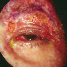
Classic herpes zoster skin rash in another patient.
The epithelial deposits were again removed and the patient was prescribed a mild topical steroid. Oral acyclovir was added at a dosage of 800mg five times per day.
When patients manifest ocular involvement from herpes virus, the most common form is herpes simplex dendritic keratitis.
The zoster virus, however, can have ocular manifestations that are often passed over or misdiagnosed because they are not as
obvious as the simplex dendritic keratitis. Without the dermatologic manifestations or neuralgia, the condition can be overlooked. This month, we discuss the other herpes.
Symptomology
Herpes zoster is caused by the varicella virus, which causes chickenpox in children.1 This virus usually enters the human system through the conjunctiva and/or nasal or oral mucosa. The body then suppresses the virus, which lies dormant in the dorsal ganglia. If the bodys immunity fails or if other triggers (such as chemotherapy or severe systemic disease) occur, the virus will actively replicate along the route of the ganglia.
Due to widespread exposure to the virus, nearly 100% of the population develops antibodies to the
disease by age 60.2 Just a small percentage experience viral activation after the initial childhood chickenpox infection. A second reactivation is not terribly common, occurring mostly in older patients with normally declining immunity and in immunosuppressed patients, such as organ transplant recipients or those who suffer from AIDS or neoplasm.2
The herpes zoster rash most
commonly resides in the facial and mid-thoracic to upper-lumbar dermatomes.1 It appears as erythematous macules and papules that typically progress into pustules within three or four days. These pustules rupture and crust over within seven to 10 days. Often, patients experience a prodrome of neuralgia several days prior to the development of dermatologic manifestations. The prodrome and dermatologic outbreak of herpes zoster can be extremely painful, and a small number of patients continue to experience post-herpetic neuralgia long after resolution of the acute outbreak.3
Ocular Impact
When the herpes zoster virus affects ocular structures, it is termed herpes zoster ophthalmicusthe virus invades and establishes residency and dormancy in the trigeminal ganglia. Upon reactivation, it travels along the ophthalmic and maxillary division of the trigeminal nerve.
Superficial ocular involvement is frequently nondescript and may manifest as punctate epitheliopathy, follicular conjunctivitis, disciform or interstitial keratitis, uveitis, scleritis or episcleritis. Epithelial necrosis can result in superficial foreign body-like lesions.
Nondescript keratitis, in the absence of neuralgia or pustular lesions along a dermatome, is often incorrectly identified as HZO. Should the keratitis coalesce into a pseudodendritic pattern, the patient is often misdiagnosed with herpes simplex keratitis. The main difference between a pseudodendritic keratitis due to HZO and true herpes simplex dendritic keratitis is the absence of terminal end-bulbs in pseudodendrites.
Herpes zoster can also cause other significant ocular manifestations that may not be commonly known as signs of the virus. Cranial nerve (especially trochlear) palsies with resultant diplopia and ophthalmoplegia can occur. Uveitis with secondary inflammatory glaucoma is also a common occurrence. Chorioretinitis as well as optic nerve involvement with subsequent vision loss also may develop.1,2,4
Therapeutic Options
Management of the ophthalmic manifestations of herpes zoster mainly involves oral antiviral agents to help suppress the virus and return it to a state of dormancy. Options include acyclovir (800mg p.o. five times a day), famciclovir (500mg p.o. t.i.d.) and valacyclovir (500mg b.i.d. to t.i.d.) for 10 days. Time is of the essence; oral therapy should be initiated within 72 hours of vesicular eruptions and ideally upon the development of the prodrome neuralgia. Early therapy is most likely to reduce the incidence of post-herpetic neuralgia.3 But, its still useful after the three-day window.
Oral steroids in conjunction with antiviral therapy may alleviate some of the discomfort in zoster outbreaks, although they are not commonly used. Mild astringents, such as calamine lotion, can be applied to the affected skin. Capsaicin cream can significantly alleviate pain when applied to the blistered skin lesions.
Also,
evidence suggests that varicella outbreaks can be prevented by vaccination. A recent trial of a varicella zoster-based vaccine (Zostavax, Merck) had a significant effect in preventing disease as well as reducing the incidence of post-herpetic neuralgia.5
While we are well aware of the classic dendritic manifestation of herpes simplex, many are less experienced when it comes to the ophthalmic clinical picture of herpes zoster. So, become familiar with the myriad occult corneal findings and other ocular manifestations of zoster, and remember to inspect for vesicular rashes and to inquire about prodrome neuralgia. By doing this, youll be prepared when you unexpectedly encounter the other herpes virus.
Post Script
Coincidentally, as this issue was going to press, a 66-year-old black woman, who had been examined and treated for tension headaches and facial swelling by a local hospital emergency room, presented because she was told to see an eye doctor and dentist for further evaluation. She reported that several days earlier, she had a painful, swollen left eyelid with a pimple, which she manually popped. It drained into her eye, causing redness. When asked, the patient indicated that her headache and facial swelling began on the top of her head and radiated to the left temporal region. She admitted that she had heat rash along the left side of her face two weeks previously.
So, did she have heat rash, a hordeolum and a tension heachace? No, she had herpes zoster.
1. McCrary ML, Severson J, Tyring SK. Varicella zoster virus J Am Acad Dermatol 1999 Jul;41(1):1-14.
2. Gurwood AS, Savochka J, Sirgany BJ. Herpes zoster ophthalmicus. Optometry 2002 May;73(5):295302.
3. Stankus SJ, Dlugopolski M, Packer D. Management of herpes zoster (shingles) and postherpetic neuralgia. Am Fam Physician 2000 Apr;61(8):2437-48.
4. Karbassi M, Raizman M, Schuman J. Herpes zoster ophthalmicus. Surv Ophthalmol 1992 May-Jun;36(6):395-410.
5. Wutzler P. Herpes zoster and postherpetic neuralgiaprevention by vaccination? Dtsch Med Wochenschr 2009 Apr;134(Suppl 2):S90-4.

