Failure to diagnose glaucoma is one of the most common causes for lawsuits against optometrists. Therefore, patients who need to be tested or treated for glaucoma must be identified. To test patients for glaucoma or glaucoma progression according to modern protocols, you might have to upgrade or acquire new equipment. This article will review which instruments are necessary for patient care and the best financial investments for your practice.
Examination Services
When you see patients for their annual comprehensive eye exam, you also perform a routine glaucoma screening. There are three billing choices for these exams. In increasing order of reimbursement and desirability, they include:
Vision plans.
Private pay.
Medically billed 92000 level ophthalmologic exams.
If you become suspicious of glaucoma while performing a comprehensive eye exam and perform additional tests, there are two billing options:
Medically billed 99000 level E/M (Evaluation and Management) exam.
Medically billed 9924X level consultation for another professional.
Because glaucoma progresses slowly, you typically do not have to perform a complete glaucoma evaluation on the same day as the comprehensive eye exam. However, tell your patients that the glaucoma tests you will perform in the future will be billed to their medical carrier rather than their vision plan.
When patients return for further glaucoma evaluation, you might use several diagnostic instruments. Each of these requires particular billing codes that provide a specific reimbursement.
|
|
| A scanning laser evaluation is now considered superior to visual field testing by some insurance carriers. |
Pachymetry
Correcting IOP readings based on central corneal thickness (CCT) measurements can help you detect glaucoma suspects. However, pachymetry can be reimbursed only once in a patients lifetime.
Some local coverage determinations (LCDs) now recognize that this restriction does not allow for the best care of glaucoma patients. For example, Noridian Medicare Services published a policy change effective May 15, 2006, that states, In those instances when more than a single pachymetry reading every two years is required (e.g., when glaucoma or ocular hypertension is initially diagnosed and one or two additional confirmatory measurements are necessary) the use of ICD-9-CM V12.49 (other disorders of the nervous system and sense organs) should be coded in addition to the primary glaucoma or corneal disease code.
Pachymeters cost about $2,000, and reimbursements average about $12 for both eyes. Therefore, youll need to take pachymeter measurements on about 167 patients before breaking even on this investment.
|
Pachymetry |
| When billing for pachymetry: Bill one unit for both eyes because pachymetry is a binocular test. Include a written interpretation and report including the corrected IOP calculation. service. |
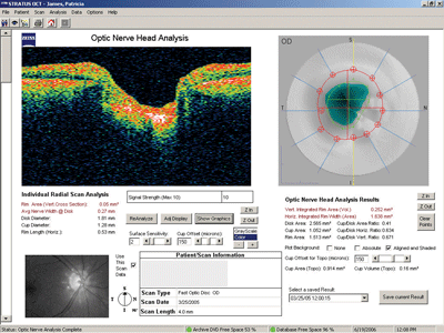
Optic nerve head analysis with a scanning laser device is superior to a single two-dimensional photo of the optic disc. Scanning laser devices can document precise cup depth, volume and shape. They have revolutionized early detection of glaucomatous optic nerve head topography. Yet, fundus photos can document other findings, such as peripapillary atrophy, which should be followed in all glaucoma patients. These images should be taken annually so progression can be evaluated. Also, fundus photos are indispensable for following disc hemorrhages.
You can bill for fundus photos for glaucoma twice a year. Typical reimbursement is about $65 to $80.
|
Fundus Photos |
| When billing for fundus photos: Bill one unit for both eyes because it is a binocular test. Include a written interpretation and report. Use CPT code 92250 to report the service. |
Scanning Lasers
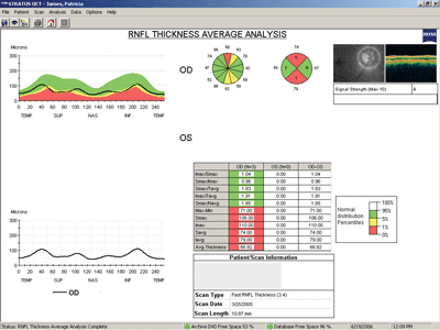
Scanning lasers have changed the way we diagnose early glaucoma. Noridians LCD for 92135 makes this clear: It (scanning laser testing) will distinguish patients with glaucomatous damage irrespective of the status of intraocular pressure.
Recently, our local carrier denied paying for scanning laser and visual field tests for glaucoma suspects when both were performed on the same visit. This is because our local carrier now considers scanning laser tests superior to visual field testing for early glaucoma detection. Their policy has been revised to state: (scanning laser testing) allows for earlier detection of optic nerve and retinal nerve fiber layer pathologic changes before there is visual field loss. Peripapillary nerve fibers must be examined because other early glaucomatous changes can remain hidden. Therefore, I no longer try to diagnose early glaucoma based on optic nerve changes, visual field tests and IOP readings alone.
Some scanning lasers can also be used to detect a variety of retinal pathologies in addition to glaucoma. Local medical policies typically permit scanning laser tests twice a year for early to moderate glaucoma. Typical reimbursement is about $40 to $50.
|
Scanning Lasers |
| When billing for scanning lasers: Bill one unit for both eyes because it is a binocular test. Include a written interpretation and report. Use CPT code 76514 to report the service. |
Visual Field Analysis
Billing for visual field tests has changed since researchers found that up to 50% of ganglion cell fibers could be lost before visual field defects appear.1,2 Emerging medical policies still recommend visual field testing to confirm early glaucoma. But, in some instances, the medical carrier will only pay for visual field tests after scanning laser analysis has detected retinal nerve fiber defects (i.e., ICD-9 code 362.85).
The frequency of visual field testing should depend on the severity of glaucoma. Two to four visual field exams per year is typical for suspect, early and moderate glaucoma. In later-stage glaucoma, visual field testing should be performed instead of scanning laser tests to monitor disease progression and efficacy of treatment. Typical reimbursement is about $65 to $80.
|
Visual Field Tests |
| When billing for visual field tests: Bill one unit for both eyes because it is a binocular test. Include a written interpretation and report. report threshold fields. |
Billing for Old and New Technology
The main purpose of performing extended ophthalmoscopy for glaucoma suspects and glaucoma patients is to binocularly evaluate the level of excavation of the optic nerve head. However, the binocular view does not allow us to detect subtle changes over time. The introduction of stereo disc photography greatly enhanced our ability to document optic nerve head topography. However, scanning laser technology has surpassed extended ophthalmoscopy and stereo disc photography in efficiency and accuracy.
Many LCDs now recognize the advanced diagnostic capability of scanning lasers. This is why many third-party payers deny payment for glaucoma workups that include more than one of the above tests. However, payment policies vary according to the local carrier, so nobody can devise a glaucoma billing protocol that is airtight in every jurisdiction.
If you determine that any of the above tests is indicated as an integral part of a patients diagnostic workup, clearly explain the need for the test in the patients record and submit it for payment. Then, review the explanation of payment (EOP) document that accompanies the reimbursement check from the insurance carrier to see which tests they reimbursed. Several of my colleagues have noted that they are more likely to be paid for multiple tests if these tests are performed on different days. So, review denials with your billing officer so you can adjust your diagnostic protocols for better reimbursements.
Medicare has a program called National Correct Coding Initiative (NCCI) to prevent clinicians from billing for procedures that they consider redundant and unnecessary. This list of mutually exclusive CPT codes can be found on their Web site under National Correct Coding Initiative (NCCI) at http://www.cms.hhs.gov/NationalCorrectCodInitEd/. The most important code pair affecting optometrists is billing for fundus photography-92250 and scanning laser-92135. This rule would likely apply for all major medical carriers. However, several O.D.s have told me they are getting paid for both of these procedures when performed on the same day and billed under various diagnostic codes. So, always bill for the procedures you consider medically necessary and review payment patterns when the reimbursement check arrives.
|
|
| Precise evaluation of optic nerve head topography in a patient with primary open-angle glaucoma. |
Keeping Up with the Times
The following technology is necessary to diagnose and manage glaucoma according to modern practice standards:
Pachymeter: $1,500 to $3,000 for single function units.
Scanning laser: $35,000 to $55,000.
Threshold visual field analyzer: $10,000 to $18,000.
Having a fundus camera for glaucoma management is a good idea, but not necessary. Fundus cameras can cost between $20,000 to $40,000. Note that the price ranges could vary considerably depending on purchasing savvy and other circumstances.
Ideally, about $80,000 will purchase the deluxe equipment package outlined above. You should earn about $1,000 in professional fees per glaucoma patient per year. If you diagnose and treat only one new glaucoma patient per month, the additional $12,000 annual revenue will take care of the payment for the above equipment. Likewise, one glaucoma patient per week can net your practice an additional $50,000 per year. Consider that any purchase scenario is cheaper than a lawsuit if you fail to diagnose glaucoma.
Owning and using the diagnostic equipment necessary for the diagnosis and management of glaucoma provides some medico-legal protection for your practice. However, solo practices often have difficulty affording equipment. Since I became part of a group practice last year, my partners and I can easily afford this equipment because we share every overhead expense. If you cannot afford the necessary equipment discussed above, consider other options, such as equipment sharing or referring your glaucoma suspects to an optometric colleague.
Dr. Dean is in a private group practice in Albuquerque, N.M. He also lectures and does on-site consulting for billing and coding.
1. Quigley HA, Addicks EM, Green WR. Optic nerve damage in human glaucoma. III. Quantitative correlation of nerve fiber loss and visual field defect in glaucoma, ischemic neuropathy, papilledema and toxic neuropathy. Arch Ophthalmol 1982 Jan;100(1):135-46.
2. Kerrigan-Baumrind LA, Quigley HA, Pease ME, et al. Number of ganglion cells in glaucoma eyes compared with threshold visual field tests in the same persons. Invest Ophthalmol Vis Sci 2000 Mar;41(3):741-8.


