Our patients come to us with more than just eye problems. Many suffer from gastroesophageal reflux disease (GERD), peptic ulcer and inflammatory bowel diseases (IBD). For this column on the digestive system, we will focus on IBD.

The Burden of Disease
An estimated 1.5 million Americans have inflammatory bowel diseases.1,2 Two IBDs are Crohn’s disease (also known as regional enteritis) and ulcerative colitis (UC).
• Crohn’s disease can involve any segment of the gastrointestinal tract from the mouth to the anus. The inflammation extends through all layers of the intestinal wall, and may involve regional lymph nodes. Cramps, tenderness, nausea, fever, diarrhea and bleeding may occur. Abscesses and fistulas may ensue, causing marked discomfort.1
Although Crohn’s disease affects both children and older adults, more than 80% of ambulatory care visits are among young and middle-aged adults. Crohn’s is most prevalent in people aged 20 to 40. It is most common among whites, and males and females are equally affected.1,2
• Ulcerative colitis, as the name suggests, is limited to the colon. UC produces edema and ulcerations. Severity may range from a mild, localized disorder to a fulminant disease that may cause a perforated colon, progressing to potentially fatal peritonitis and toxemia.1
In 2004, there were about one-half million first-listed-diagnosis ambulatory care visits for UC and about 700,000 all-listed visits. Visit rates were highest among young adults, and women had almost twice the rate of men. Patients with UC have an increased risk of getting colorectal cancer. Children may experience impaired growth.1-3

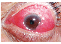
Scleritis and other ocular inflammations may precede a diagnosis of ulcerative colitis or Crohn’s disease.
First-degree relatives have a five- to 20-fold increased risk of developing IBD compared with those in unaffected families. The child of a parent with IBD has a 5% risk of developing IBD. Twin studies show a concordance of approximately 70% in identical twins vs. 5% to 10% in non-identical twins.3
Theories of Pathogenesis
The causes of IBD are currently unknown. It is thought that the etiologic process involves an immune reaction of the body to its own intestinal tract. Activation of the immune system leads to inflammation of the intestinal tract, both acute and chronic. The common end pathway is inflammation of the mucosal lining, causing ulceration, edema, bleeding, and fluid and electrolyte loss.
Persons with IBD may have a genetic predisposition for the disease. The triggering event for the activation of the immune response has yet to be identified. Possible factors related to this event include a pathogenic organism (such as Escherichia coli), an immune response to an intraluminal antigen (such as protein from cow’s milk) or an autoimmune process.1,3
Disease Complications
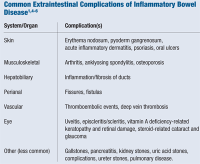
The most typical manifestation of UC is bloody diarrhea. Pain is uncommon, but may occur. The most typical manifestations of Crohn’s disease are abdominal pain and diarrhea. Malabsorption secondary to IBD may result in deficiency of vitamin A, the B vitamins and folate. In both types of IBD, patients may demonstrate marked weight loss and weakness; anemia frequently accompanies disease activity.3,4 (See “Common Extraintestinal Complications of Inflammatory Bowel Disease”)
Ophthalmic complications occur in approximately 3% of patients with IBD, and are more frequent in UC than in Crohn’s disease. The major conditions involved are episcleritis, scleritis and uveitis.4,5 In addition to local anti-inflammatory therapy, these ocular conditions usually respond well to treatment of the underlying bowel disease.6

Diagnosis and Treatment
Serology can help detect such changes as anemia, high white blood cell counts (indicating inflammation or infection) and low nutrient levels. Stool samples can rule out intestinal infections, which may lead to similar symptoms as those of IBD.1
The most direct way to make a firm diagnosis of IBD is by endoscopy, biopsy or barium X-ray.1,7 With endoscopy, the lining of the intestinal tract can be observed and biopsies can be obtained. An abdominal CT scan or MRI may be useful to detect abdominal abscesses in patients with Crohn’s disease. Capsule endoscopy is a newer test in which the patient swallows a pill-sized camera, which travels through the small intestine taking pictures that are transmitted to a recorder and later viewed on a computer interface.1,7
The goals of IBD treatment are to control inflammation, replace nutritional losses and blood volume, and prevent complications. Management may include bed rest, I.V. fluid replacement, vitamin B12 injections, elimination of dairy products and a clear-liquid diet. If anemia is present, blood transfusions or iron supplementation may be needed. Surgery may ultimately be indicated to correct for bowel perforation, hemorrhage, fistulas and intestinal obsruction.
Because UC and Crohn’s disease are chronic illnesses, they often require long-term treatment with medications. Aminosalicylates are among the most commonly used drugs to treat IBD and include agents such as sulfasalazine (Azulfidine [Pfizer]), mesalamine (Asacol [Warner Chilcott], Pentasa [Shire] and Lialda [Shire]) and balsalazide disodium (Colazol [Salix]). The active component of these medications is 5-aminosalicylic acid, which works to reduce inflammation in the intestinal wall.
Immunomodulatory therapies, including corticosteroids, 6-mercaptopurine (Purinethol [Teva GTC]), azathioprine (Imuran [Prometheus Labs]) and methotrexate work to bring active disease under control, and keep IBD in remission.1,7,8

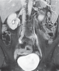
MRI with contrast shows enhancement of thickened bowel wall in inflammatory bowel disease.
Eye-related complaints and signs may precede a diagnosis of UC or Crohn’s disease. Because many patients are unaware that IBD has a risk of ocular complications, patient education is vital. And, ocular health evaluation should be a routine component in the care of patients with IBD. An integrated, multidisciplinary approach to the care of these patients can be helpful in optimizing management.
1. Gastrointestinal Disorders (chapter 4). In: Professional Guide to Diseases, 9th ed. Philadelphia: Wolters Kluwer/Lippincott Williams & Wilkins; 2009: 233-98.
2. Everhart JE (ed.). The burden of digestive diseases in the United States. US Department of Health and Human Services, Public Health Service, National Institutes of Health, National Institute of Diabetes and Digestive and Kidney Diseases. Washington, DC: US Government Printing Office, 2008; NIH Publication No. 09-6443;97-102.
3. Andres PG, Friedman LS. Epidemiology and the natural course of inflammatory bowel disease. Gastroenterol Clin North Am. 1999 Jun;28(2):255-81, vii.
4. Rothfuss KS, Stange EF, Herrlinger KR. Extraintestinal manifestations and complications in inflammatory bowel diseases. World J Gastroenterol. 2006;12:4819-31.
5. Khan A, Lightman S. The eye in gastrointestinal disease. Hosp Med. 2003 Sep;64(9):548-51.
6. Mintz R, Feller ER, Bahr RL, Shah SA. Ocular manifestations of inflammatory bowel disease. Inflamm Bowel Dis. 2004;10:135-9.
7. American Society for Gastrointestinal Endoscopy. The role of colonoscopy in the management of patients with inflammatory bowel disease. Gastrointest Endosc. Dec 1998;48(6):689-90.
8. Kozuch PL, Hanauer SB. Treatment of inflammatory bowel disease: A review of medical therapy. World J Gastroenterol. 2008;14:354-77.

