 |
I (Dr. Kabat) must have burned my tongue on something. That was the only logical explanation. I was eating a delicious dinner on my 54th birthday, but it didn’t taste right. In fact, it barely tasted at all. Maybe some more salt? Nope…well, this is depressing. It’ll be better tomorrow, I thought.
Breakfast the next morning was equally bland. At work, I noticed that my right eye was tearing excessively, and that was unusual. Did I injure it somehow? Well, the eye felt a little bit scratchy. Some artificial tears should take care of it. Unfortunately, it kept on tearing throughout the day, and my eye felt… funny. Not painful, but almost like I had put a drop of tetracaine in my eye. Boy, this really isn’t my week.
It wasn’t until I started shaving on the third day that I realized what was going on. As I tried to puff out my cheeks, I found myself sputtering and spitting on the mirror. I looked closer at my face. I smiled a wide grin, and to my astonishment, only the left side of my face responded.
 |
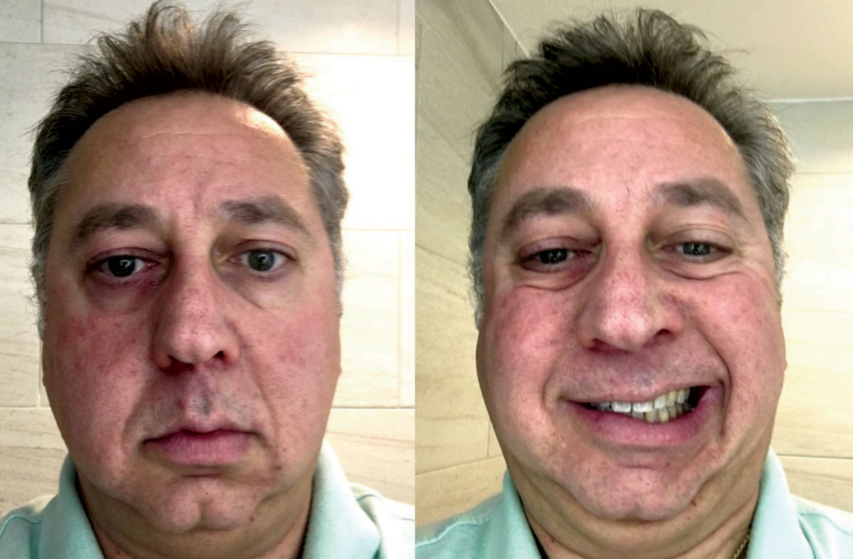
At left, note the flattening of the right side of the face, with drooping of the nasolabial fold, corner of the mouth and lower eyelid. At right, when smiling, the teeth remain unexposed on the affected right side. |
OK, think. You’re a doctor, after all. Are you having a stroke? I quickly checked my motor function. Both arms and legs seemed to be working alright. Memory? I knew my name, my address, where I was and where I was going that day. I thought, ‘Let’s run through cranial nerves.’ CN I (olfactory), check. CN II (optic), vision was fine in both eyes and no apparent hemispheric field loss. CN III, IV and VI, no diplopia in any gaze. CN V (trigeminal), sensation on both sides of the face are equal and muscles of mastication are working fine. CN VII (facial), definite disparity on the right side. My blink appeared asymmetric, favoring the left side, although I could squeeze the right eye shut if I tried. So my orbicularis oculi, orbicularis oris and buccinator function were all compromised on the right side. CN VIII (vestibuloauditory), no balance problems and I seemed to be hearing equally in both ears. CN IX and X, no problems swallowing or coughing (but I refused to check my own gag reflex). CN XI, shoulder shrug and neck turns were equal to both sides. CN XII, stuck out my tongue and it was straight. Phew!
What's My Problem?
So, what’s my diagnosis, doc? If you thought “Bell’s palsy,” then we’re on the same page. Bell’s palsy represents an idiopathic dysfunction of CN VII, and is the most common presentation of facial nerve palsy.1-4 The characteristic clinical presentation involves generalized weakness of one side of the face. There will be an inability to fully close the ipsilateral eye, which can result in lower lid ectropion and epiphora; with persistent lagophthalmos, patients may manifest conjunctival hyperemia and exposure keratopathy, resulting in dry eye symptoms. Additionally, there will be unilateral flattening of the nasolabial fold, drooping of the corner of the mouth and diminished wrinkling of the forehead. These signs become more evident when asking the patient to purse the lips, puff out the cheeks, smile widely and raise the eyebrows. Since the CN VII also supplies sensory innervation to the anterior two-thirds of the tongue, altered or decreased taste sensation is common. Depending upon the severity and branches involved, patients may additionally report pain in or behind the ear, as well as hyperacusis—increased sensitivity to sound—on the affected side.
The onset of all these symptoms is typically abrupt, although, as in my case, it may be several days before the patient fully recognizes the magnitude of their disability. By the time a patient presents for evaluation, it is likely that 24 to 72 hours have already passed.
For Whom the Bell's Tolls
The literature shows some debate regarding the epidemiology of Bell’s palsy. The reported annual incidence varies throughout the world, with estimates varying between 11 and 40 cases per 100,000 individuals.3,5 It has no known gender, ethnic or racial predilection. Most cases are seen in mid- to later-life and the median age of onset is 40.1-4,6-9 Known risk factors include diabetes, pregnancy, severe preeclampsia, obesity and hypertension.2-4 The underlying pathophysiology of Bell’s palsy, as observed in post-mortem cases, involves vascular distension, inflammation and edema with associated ischemia of the facial nerve.5
As to the etiology, the condition is classified as idiopathic, but current thinking suggests that it is most likely associated with reactivation of herpes simplex virus (HSV) or herpes zoster virus (HZV) from the geniculate ganglia.2,5
Although the clinical presentation may be easily recognized, physicians need to always bear in mind that Bell’s palsy is a diagnosis of exclusion. Despite wanting to spare our patients the time and expense involved, a thorough medical evaluation should be obtained in these scenarios. Approximately half of all facial nerve palsies are idiopathic and fall into the category of Bell’s palsy, but that means 50% have another cause.
Acquired facial nerve palsies may be associated with trauma, ischemia, systemic infection (e.g., Lyme disease or tuberculosis), granulomatous disorders (e.g., sarcoidosis or granulomatosis with polyangiitis), autoimmune disease, vasculitis, numerous viruses (e.g., coxsackievirus, cytomegalovirus, adenovirus, mumps, rubella, influenza B and Epstein-Barr), or even neoplastic disorders. Laboratory testing and radiologic evaluation are essential to rule out these other potential causes, or any substantial comorbidities that may have been undiagnosed previously. While a “shotgun” diagnostic approach is discouraged in these cases, a targeted systemic workup based on the patient’s personal and family history as well as concurrent symptoms or signs is essential.
Comanagement with the patient’s primary care physician may be the best and most efficient way to obtain this information. In my case, MRI with and without contrast of the brain was ordered as a prophylactic measure.
Unringing that Bell
Strong evidence suggests that systemic corticosteroids may hasten recovery of Bell’s palsy.8,10-14 The preferred therapeutic regimen is prednisolone 60mg/d (in divided doses) for five days, then subsequently tapered for five additional days.13 Ideally, treatment should be initiated within 72 hours following the onset of symptoms.8,10-14There is less agreement among experts regarding the role of antivirals in acute Bell’s palsy. Some studies show a modest benefit to oral antivirals, but the prevailing opinion is that these medications alone are no better than placebo. If considered at all, oral antiviral agents (e.g., valacyclovir 1,000mg TID for seven days) should be given in conjunction with oral corticosteroids, but only after alternate infectious causes have been eliminated.8,10-17 Non-traditional, but potentially beneficial, therapies may include acupuncture, hyperbaric oxygen therapy and various forms of physical therapy.18-21
The primary optometric goal in Bell’s palsy is mitigating the effects of exposure keratopathy. Lubricating drops or ointments, or both, can be helpful for symptoms, but a bandage contact lens may be more protective to the cornea and provide greater, lasting relief. Nighttime exposure due to lagophthalmos can be prevented by taping the affected lids closed or by using a sleep/moisture retention mask. I found the Eyeseals Hydrating Sleep Mask (Eye Eco) to be exceptional. The use of external eyelid weights, such as Blinkeze (MedDev Corporation) should be considered in the elderly, those with diabetes and those with pre-existing eye disease.22
Many individuals with Bell’s palsy will recover fully within several weeks to months, although some potential facial paralysis may linger. In such cases, surgical intervention may be a consideration, although the evidence for success is not extensive.4,23 Failure to observe resolution of signs after three months should prompt the clinician to consider an alternative diagnosis and initiate additional medical testing.
As I write this, it has been 10 days since my initial onset of symptoms, and I’m already noting recovery of both sensory and motor function. I consider myself fortunate. I’m even curious to see what the inside of an MRI machine looks like.
Taking it to the Limit
On a very different note, I want to let our regular (and occasional) readers know that I will no longer be a regular contributor to Therapeutic Review. It has, in fact, been 14 years since Dr. Sowka and I penned our first column, and in the immortal words of Harry Callahan (Clint Eastwood), “A man’s got to know his limitations.” I am honored to have had the opportunity to share my clinical experience and insight with my colleagues and will continue to do so in other venues and media. Thanks to our numerous editors over the years for their talent, encouragement and patience, especially Mike Hoster and Bill Kekevian.
|

