 History
A six-year-old white male presented emergently with a chief complaint of a watery, diffusely red left eye that had persisted for four days. His mother explained that she had taken the boy to his pediatrician and received some antibiotic eye drops; however, she confessed to prematurely discontinuing the regimen after five days because it appeared that the agent was not working effectively.
Otherwise, the child had unremarkable ocular and systemic histories. Neither the patient, nor his mother, reported any known allergies.
Diagnostic Data
His best-uncorrected entering visual acuity measured 20/20 OD and 20/30 OS at distance, and 20/20 OU at near. External examination was normal, with no evidence of afferent pupillary defect OU. Refraction revealed symmetrical, spherical hyperopia of +0.50D, and yielded no improvement in visual acuity.
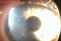
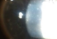
The anterior segment evaluation of the right eye was unremarkable; however, the left eye exhibited significant findings (figures 1 and 2).
The patient had difficulty keeping his eyes open during applanation tonometry, which required us to forcibly squeeze open his lids. Eventually, we were able to secure an intraocular pressure measurement of 19mm Hg OU.
The dilated fundus examination of both eyes was unremarkable, with a cup-to-disc ratio of 0.30 x 0.30 OU. Further, he had distinct rims, pink discs, normal maculae and no peripheral pathology OU.
Your Diagnosis
How would you approach this case? Does the patient require any additional tests? What is your diagnosis? How would you manage this patient? What is the likely prognosis?
Discussion
Additional testing should include an expanded medical history to rule out a previous or recent upper respiratory infection. Sodium fluorescein instillation or lissamine green dye could be used to rule out keratitis or focal corneal compromise, and rose bengal dye might be instilled to assess the cornea for devitalized lesions. Further, a cotton-tipped wisp could be used to determine and compare corneal sensation in both eyes.
Be sure to evert both the top and bottom eyelids to rule out the presence of pseudomembrane formation. Also, inspect the lids for evidence of mucopurulent discharge, follicles and/or papillae. Finally, in all cases of red eye, a cursory inspection of the lymph nodes should be completed to rule out lymphadenopathy.

|
 |
|
This six-year-old boy presented with a red, watery left eye (OD left, OS right). What do you notice, and how should he be treated?
| |
Because the patient’s eye was diffusely red without mucopurulent discharge, the conjunctiva was dotted with follicles, his cornea was clear and the topical antibiotic had no effect after five days of use, we diagnosed him with epidemic keratoconjunctivitis (EKC).
Viral conjunctivitis may be caused by a number of different infectious agents. In most instances, the ocular sequelae are both mild and self-limiting; however, the potential exits for a severe, prolonged, debilitating episode with ocular discomfort and visual disability.1-6 The two most frequently encountered forms of self-limiting viral conjunctivitis are EKC and pharyngoconjunctival fever.1-6
• Epidemic keratoconjunctivitis presents as a bilateral, inferior palpebral, follicular conjunctivitis with epithelial keratitis and subepithelial infiltration. Corneal sensation is unaffected. The subepithelial infiltrates are typically concentrated in the central cornea, sparing the periphery. EKC is regularly caused by adenovirus types 8 and 19.1-3,5,6
• Pharyngoconjunctival fever is characterized by a triad of fever, sore throat and follicular conjunctivitis. It may be unilateral or bilateral. The condition is predominantly caused by the adenovirus type 3, but may occasionally result from subspecies 4 and 7.1,3,5,6 The disorder varies in severity and can persist between four days and two weeks.1-6 While the virus may be eradicated from the conjunctiva in 14 days, it can remain active in fecal excretions for up to 30 days. This may explain why epidemics are often traced to swimming pools.
In both instances, the associated signs and symptoms of conjunctival injection–tearing, watery discharge, red edematous eyelids, pinpoint subconjunctival hemorrhages, pseudomembrane (with occasional true membrane) formation and palpable preauricular lymphadenopathy–often vary in severity and frequently mimic those caused by bacterial conjunctivitis.1-5
Viral/follicular conjunctivitis appears to be the result of a host response to an exogenous substance. The disease is spread either by an airborne vector or via direct transfer from fingers to the conjunctival eyelid surface.1-6 After an incubation period of five to 12 days, the disease enters the acute phase, which includes a watery discharge, conjunctival hyperemia and follicle formation.3 Lymphoid follicles are elevated, avascular lesions ranging from 0.2mm to 2.0mm in size that consist of germinal centers.
The confounding issue is that this type of conjunctivitis often does not present in the classic manner, leaving practitioners to speculate on a presumed diagnosis, rather than react to a positively identified cause. Hence, eye care providers frequently attempt an empiric, broad-spectrum approach to nebulous red eye disease by prescribing a topical antibiotic or a combination antibiotic/steroid. Other topical medications include nonsteroidal anti-inflammatory medications or combination mast cell stabilizers/antihistimines.5
Some may argue that there is little harm in using these topical agents in concert, but clearly antibiotics play no role in resolution of ocular adenoviral illness. In fact, these medications can produce corneal toxicity and a variety of side effects, are an unnecessary expense and may be tremendously inconvenient to administer, particularly in pediatric patients. Further, if the patient is not positively diagnosed with viral conjunctivitis—and thus not advised of his or her highly contagious status—the individual may return to work or school, potentially spreading the condition to others.
Instead of a purely empiric evaluation, consider testing the patient’s tear film with the AdenoPlus (Nicox), an in-office device that may more easily facilitate a diagnosis.6 Its sample collector is designed to safely capture and transfer an appropriate ocular fluid sample from the lower conjunctiva to the immunoassay, which is located in a plastic cassette. Once the sample has been transferred, the result is ready in approximately 10 minutes, very similar to an at-home pregnancy test.
Because viral conjunctivitis is contagious and self-limiting, the primary function of ophthalmic management is to increase the patient’s awareness by providing education and ensuring comfort by decreasing symptomatology with topical agents and/or supportive therapies.
Anecdotally, povidone-iodine antimicrobial solution has been advocated as a therapeutic measure for patients in severe discomfort. Clinical research has now added some support for this treatment. Within the last two years, researchers analyzed the use of 2% povidone-iodine (PVP-I) solution on EKC.7 A prospective, interventional, uncontrolled study of 61 EKC patients evaluated the clinical effects of QID PVP-I therapy.7 The researchers concluded that topical application of 2% PVP-I was both tolerable and effective for relieving ocular discomfort secondary to EKC.7
We educated the patient’s parents about the contagious nature of his condition, and recommended that they keep the child out of school as long as his eye remained red. Because the topical antibiotic already was initiated, we decided to continue it for the balance of seven days before complete cessation.
We recommended artificial tears and cold compresses PRN to increase comfort. Because the cornea was not involved, we prescribed a topical non-steroidal anti-inflammatory medication QID OS. Then, we informed the patients that these infections typically persist for two full weeks before resolution begins. Finally, we scheduled him for a 10-day follow-up examination.
At the follow-up, we noted that resolution had already begun. Over the next two weeks, the condition fully resolved, yielding a final visual acuity of 20/20 OU.
1. Vaughan D, Asbury T. Conjunctiva. In: Vaughan D, Asbury T. General Ophthalmology, 10th ed. Los Altos, Calif.: Lange Medical Publications; 1983:58-84.
2. Cullom RD, Chang B. Conjunctiva/Sclera/External Disease: Viral Conjunctivitis. In: Cullom RD, Chang B. The Wills Eye Manual: Office and Emergency Room Diagnosis and Treatment of Eye Disease. Philadelphia: J.B. Lippincott Co.; 1994:110-1.
3. Dawson CR, Sheppard JD. Follicular Conjunctivitis: In: Duane’s Clinical Ophthalmology. Philadelphia: J.B. Lippincott Co.; 1995:1-20.
4. Gigliotti F. Acute conjunctivitis. Pediatr Rev. 1995 Jun;16(6):203-7.
5. Kinchington PR, Romanowski RG, Jerold Gordon Y. Prospects for adenovirus antivirals. J Antimicrob Chemother. 2005 Apr;55(4):424-9.
6. Rapid Pathogen Screening. Point-of-care diagnostic services. Adeno Detector instructional insert. RPS Insert; 2006:1-2.
7. Trinavarat A, Atchaneeyasakul LO. Treatment of epidemic keratoconjunctivitis with 2% povidone-iodine: a pilot study. J Ocul Pharmacol Ther. 2012 Feb;28(1):53-8.

