 |
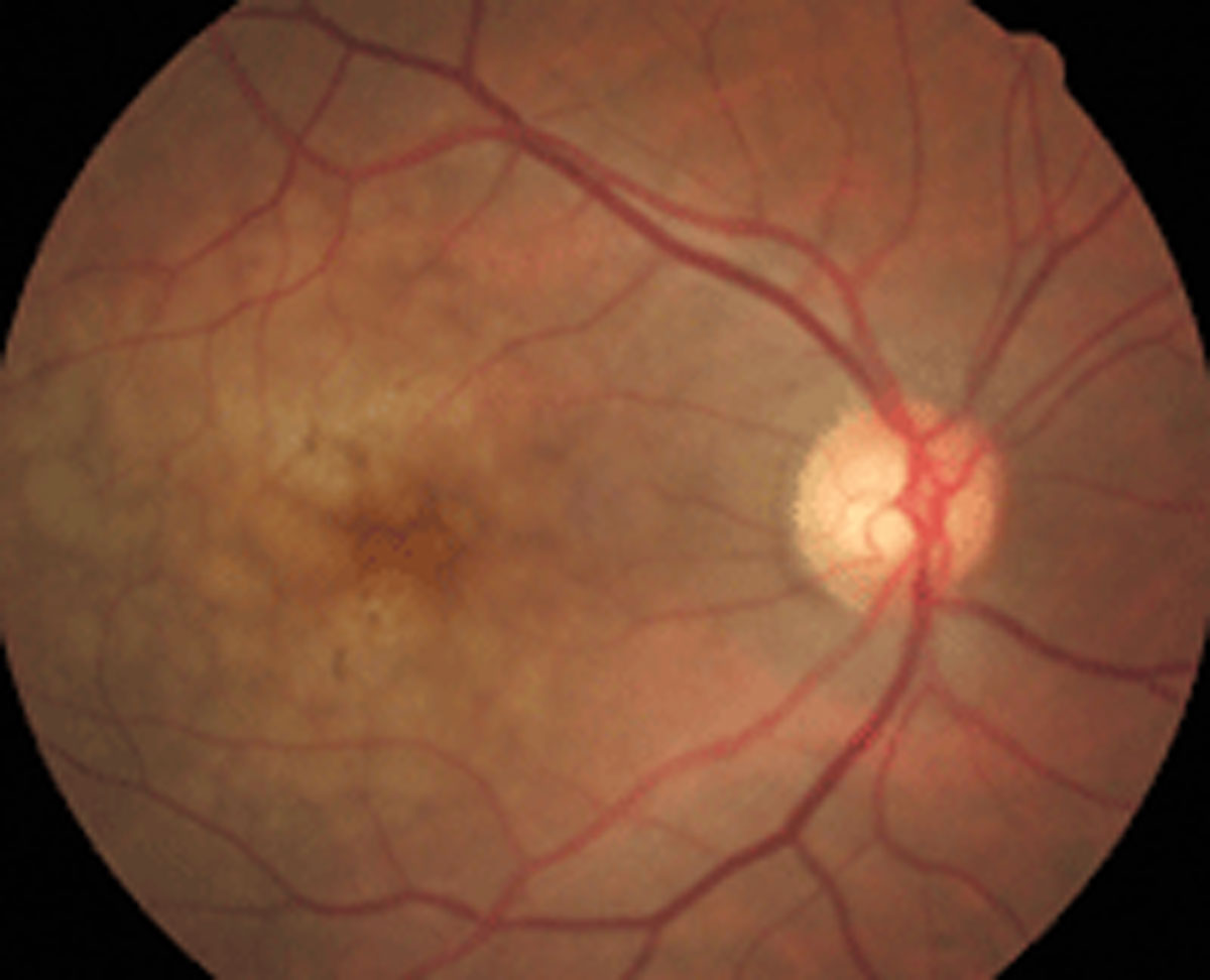
New imaging biomarkers aid in AMD detection and treatment. Photo: Diana L. Shechtman, OD, and Paul M. Karpecki, OD. Click image to enlarge. |
Patients with neovascular age-related macular degeneration (nAMD) often have subretinal fibrosis (SF) despite regular control visits and standardized anti-VEGF treatment. SF causes irreversible retinal damage, but it’s difficult to detect and quantify on standard imaging modalities. Researchers in Vienna recently evaluated the morphologic and microvascular differences between eyes with and without SF due to neovascular AMD using standard imaging and swept-source OCT angiography. They found that a multimodal imaging approach can detect important differences.
The prospective, cross-sectional study included 60 patients (37 female) with nAMD and a minimum history of 12 months of anti-VEGF treatment. A total of 20 (33%) were diagnosed with SF. The researchers reported that SF eyes had a significantly higher prevalence of outer retinal tubulation (ORT) and lower prevalence of hyperreflective foci than eyes without SF.
Of the 50 eyes analyzed quantitatively for microvascular biomarkers, the researchers reported that eyes with SF had a larger greatest vascular caliber and greatest linear diameter, larger microvascular neovascularization (MNV) area, larger vessel area, higher number of vessel junctions, longer total vessel length, higher number of vessel endpoints and higher endpoint density.
The researchers concluded that using multimodal imaging “demonstrated in vivo microvascular and morphological differences in eyes with MNV secondary to nAMD with and without SF.” They noted that eyes with SF tended to have larger MNV lesions with thicker vessels, which were often associated with the presence of ORT.
Roberts PK, Schranz M, Motschi A, et al. Morphologic and microvascular differences between macular neovascularization with and without subretinal fibrosis. Transl Vis Sci Technol. 2021;10(14):1. |

