 |
Q:
I saw a patient this morning who had fallen a week ago, hitting directly on the back of his head. One day later, he presented to the ER with vertical diplopia. They ordered a computed tomography (CT) scan, which was normal. What’s at the root of the symptoms?
A:
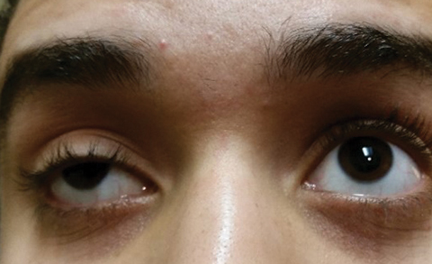
One day after the patient fell, he noticed the double vision, which went away with closing either eye, says Dennis Mathews, OD, of Eye Specialty Group in Memphis, Tenn. “With the exception of a two to three prism diopter left hypertropia, distance and near, his eye exam was normal this morning. My first impression: The left superior oblique muscle is the culprit,” says Dr. Mathews.
 |
| Besides trauma, myasthenia gravis should be ruled out in everyone with acquired vertical diplopia. |
Rationale and Differential
The fourth cranial nerve, also called the trochlear nerve, originates in the dorsal midbrain. “The nerve is long and thin and courses along the tentorum, petrosal ridge and the sphenoid ridge. It is highly sensitive to closed head trauma with small hemorrhages possible,” says Dr. Mathews.
Traumatic fourth nerve palsies may be bilateral in a minority of cases, but are usually unilateral, explains Dr. Mathews. “Skew deviations may look like fourth nerve palsies, but these lesions do not show a torsional component, may be comitant early and show other brainstem or cerebellar signs,” he says. Those include lower brainstem signs, such as internuclear ophthalmoplegia, and coordinated motor defects if the cerebellum is involved.
The differential includes a cavernous sinus lesion, which was ruled out by motility exam, as these normally are associated with oculomotor nerve palsy, abducens nerve and Horner’s pupil. Other possible causes are tumor, infection, aneurysm, diabetes and multiple sclerosis. With cranial nerve palsies, practitioners should always consider myasthenia gravis when evaluating acquired vertical diplopia in adults and children.
“In this case, with the history that was obtained, traumatic trochlear nerve palsy is the correct diagnosis. The anterior medullary velum is a common place for a traumatic lesion to present. This area is located under the fourth ventricle in the caudal dorsal membrane.”
Pinpointing the Nerve
Using the Park three-step test can allow practitioners to differentiate the nerve responsible for causing the vertical defect, says Dr. Mathews. “After the side of hypertropia is identified, the patient is asked to look to both sides with the hyper deviation being compared. If the hyper deviation increases when looking toward the opposite side, a superior muscle is involved; otherwise, an inferior muscle is involved.”
The four possible muscles causing our patient’s problem are the left superior oblique, the left inferior rectus, the right superior rectus and the right inferior oblique. “In this patient’s case, the three-step test showed the left superior oblique was involved. While the brain CT scan was normal, these images are superior to magnetic resonance imaging (MRI) only in revealing acute blood or bony defects, but not soft tissue disease,” says Dr. Mathews. He adds that an MRI would be appropriate to discover conditions such as multiple sclerosis, a tumor in the cavernous sinus or brain stroke. Magnetic resonance angiography would be used to discover an aneurysm.
Treatment
Dr. Mathews recommends patching to get the patient through the next month. One unobtrusive way to cover the involved eye is with a few pieces of scotch tape on the back surface of the patient’s glasses.
Dr. Mathews says that, even though a general timeline doesn’t exist to consider bloodwork, imaging, prism and, ultimately, surgery if the patient doesn’t improve after one month, consider referral to a neuro-optometrist, neuro-ophthalmologist or neurologist for a second opinion.

