Decades ago, eye care providers didn’t have many over-the-counter (OTC) dry eye treatments, let alone pharmaceutical or surgical options. That’s not the case today, however. Now, a plethora of viable treatments are available for dry eye and meibomian gland dysfunction patients.
This contemporary surge in effective choices largely is due to an improved understanding of ocular surface disease and patient care.
The multifactorial etiology of dry eye warrants a detailed diagnostic approach. Thus, the prudent management course may not involve ocular surface treatments at all, but rather environmental, functional or pharmacological modifications. Focused and individualized therapy should only be determined following a thorough history and in-office work-up.
Conventional Treatments
Lid scrubs, compresses and digital massage still comprise the first line of dry eye treatment. Single-use lid scrubs offer convenience to those who desire an alternative to traditional methods, such as cleansing with baby shampoo. Hot compresses should be prescribed with the awareness that meibomian secretions from severely obstructed glands possess a higher melting point.1 Digital lid massage effectively targets the meibomian glands, but may cause warping if performed over a heated cornea.2
OTC: “Oh, the Choices”
Many dry eye patients self-treat before seeking professional help.3 And given a choice, cost-conscious dry eye sufferers likely will lean toward less expensive, store-brand, artificial tears. Although generally well tolerated when dosed up to six times per day, these BAK-preserved lubricants can lead to corneal toxicity and increased inflammation.4,5 Prescribing beyond this interval, however, merits a switch to either a preservative-free option or one that breaks down upon ocular surface contact (e.g., sodium perborate). However, increasing the viscosity will permit a less frequent dosing schedule.
As evaporative dry eye accounts for the majority of cases, lipid-based products can provide symptomatic relief in many (though not necessarily all) patients.6 These include FreshKote (Focus Laboratories), Refresh Optive Advanced (Allergan), Retaine MGD (Ocusoft), Systane Balance (Alcon) and Soothe XP (Bausch + Lomb). Specific brand selection generally is left up to the eye care provider’s discretion. Single-dose, unpreserved artificial tears also are also available, such as Refresh Optive Sensitive (Allergan).
Fish oil supplementation is a potential alternative to topical dry eye treatments. Long-chain omega-3 fatty acids (eicosapentaenoic acid [EPA] and docosahexaenoic acid [DHA]) present in fish oil capsules provide an anti-inflammatory effect, which often helps improve the signs and symptoms of meibomian gland dysfunction (MGD).7 Common side effects of fish oil supplementation include gastrointestinal complications and “fishy” burps.

|
|
|
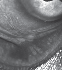
On infrared meibography, gland dropout appears as dark areas vs. light areas in meiboscopy. Glands exhibit multiple morphological changes, which may include dilation, truncation or complete atrophy.
|
To complement any of the aforementioned dry eye treatments, you might consider the use of moisture goggles. Some technologies, such as Tranquileyes (Eyeeco), have been specifically designed for dry eye treatment; however, simple swimming goggles may be used, as well.8
Stick to the Script
In recent years, optometrists have written more prescriptions for topical steroids and non-steroidal inflammatory agents than ever before.9 And, without question, underlying inflammation is one of the primary causes of dry eye.10
When considering prescription treatments for dry eye, most eye care providers start with Restasis (cyclosporine, Allergan) BID in conjunction with a topical steroid for several weeks or months to rapidly suppress inflammation (see “A Word About Steroids,” below). After a year of Restasis treatment, dosing may be reduced to QD in order to maintain a therapeutic benefit.11
The broad-spectrum topical macrolide, azithromycin, also has been shown to decrease inflammation and improve lipid alteration in patients with dry eye disease and meibomian gland dysfunction (MGD).12,13 A four-week course of the sustained delivery drop, dosed BID for two days then once daily, may effectively improve foreign body sensation and meibomian gland obstruction.14
Tetracyclines offer an oral alternative to inhibit meibomian gland inflammation.12 Of these agents, doxycycline appears to be the common selection.12 Dosing varies depending upon presentation severity, and may be as high as 100mg BID (although doses as low as 40mg QD can be effective).15 When prescribing tetracyclines, be sure to educate patients about the potential effect on oral contraception.
Secretagogues represent an additional oral treatment for dry eye, though with undesirable parasympathomimetic side effects. In a systematic review of published trials, both cevimeline and pilocarpine demonstrated symptomatic improvement in dry eye.16
Outside of topical and oral treatments, once-a-day Lacrisert (hydroxypropyl cellulose ophthalmic insert, Valeant) may be a potential option. These inserts work well for dry eye patients who have mental or physical conditions that prohibit topical drop instillation, or if convenience is a priority.17
Should the aforementioned therapeutic options fail, compounded topical tacrolimus––an immunosuppressive agent used after organ transplant––has been shown to improve tear stability and ocular surface status.18 It is also available as an ointment, making overnight coverage possible.
Additionally, compounded 3% testosterone cream applied at bedtime has been shown to increase tear osmolarity and improve evaporative dry eye.19,20 Exogenous testosterone use, however, may be an independent risk factor for central serous chorioretinopathy.20
 | |
|
Lissamine green staining on the line of Marx. An irregular, thickened or anteriorly placed line of Marx can produce ocular surface symptoms and could serve as an indication for debridement-scaling technique.
|
Plugs, Probes and Power Tools
When punctal plugs first gained popularity more than 20 years ago, many of us simply used the devices when patients complained of dryness. However, today we know that any inflammatory component must be addressed before punctal plugs may be considered. Also, depending upon the type and size of plug selected, extrusion may occur.
Before the widespread acceptance of punctal plugs, thermal cautery of the puncta was a frequently employed treatment option for recurrent dry eye. A study published in 1998 indicated that this technique yielded better subjective improvement in dry eye symptoms than argon laser punctal ablation.21 To minimize tissue destruction, some surgeons prefer a low-temperature/high-frequency cautery device.22
For meibomian glands, the debridement-scaling technique promoted by Donald Korb, OD, and Caroline Blackie, OD, PhD, may be used to remove accumulated tissue and debris from the line of Marx (LOM) and keratinized lid margin.23 To accomplish this, apply lissamine green dye and use a lateral motion with the golf club spud along the LOM to remove the stained cells.
If the meibomian glands are obstructed, several methods exist. Intraductal probing, for example, allows the eye care provider to penetrate and clear the meibomian orifice. First described by ophthalmologist Steven L. Maskin, this technique yields symptomatic improvement in all patients tested.24,25
Additionally, an automated device, the LipiFlow Thermal Pulsation System (TearScience), could be considered. The technology includes a lid warmer (which resembles a larger scleral lens) that heats to 42.5°C and an inflatable/deflatable cup that rests over top of the closed eyelids and “milks” the meibomian glands. One study suggested that the LipiFlow system improved both signs and symptoms of dry in after 12 minutes of use.26
Patient, Heal Thyself
Severe dry eye or persistent epithelial defects may warrant the use of autologous serum (AS) drops. This preservative-free, 20% to 100% dilution is formulated using the patient’s own nutrient-rich blood (see “How and Why to Make Autologous Serum,” March 2012). These drops contain growth factors, fibronectin and vitamins that help to promote ocular surface healing.27,28
AS drops generally are dispensed in single-use vials, which can be preserved for at least one month if kept refrigerated and three months if kept frozen. The typical dosing frequency of AS ranges from Q1H to QID, depending upon disease severity.27
Take note that patients with HIV, hepatitis C or syphilis are not candidates for AS formulation, and instead must use ready-made, ABO-matched serum.29
Additionally, commercially available amniotic membrane grafts also promote ocular surface healing via anti-scarring, antimicrobial and anti-inflammatory effects.30 Practitioners may choose choose from three different graft sizes, depending on the level of inflammation. They also may be employed after conjunctivochalasis surgery to prevent tear meniscus changes and delayed tear clearence.31,32
Today, eye care providers are in an excellent position to treat dry eye disease. Research into novel, even more advanced treatments remains ongoing, and new options will continue to trickle down the pipeline.
However, finding the right treatment––or combination of treatments––remains the key to successful, individualized dry eye care.
|
Stop, Look and Listen Pinpointing the underlying cause of dry eye will help you most effectively manage patient symptoms. Here’s how to do it: • History. To start, take a detailed history. Assess functional issues (e.g., prolonged reading, computer use and monitor height), visual fluctuations, environmental factors (e.g., working conditions, smoking status, driving commute duration, use of household humidifiers, contact lens wear, ceiling fan use), current medications and any history of ocular surgery or trauma. For contact lens patients, document both wearing schedules and the solutions used. Also, be sure to offer dry eye questionnaires, such as the Ocular Surface Disease Index (OSDI) or the Standard Patient Evaluation of Eye Dryness (SPEED, TearScience). • Testing. Next, perform a variety of diagnostic tests. Start with fluorescein dye instillation to assess tear film break-up time (TFBUT), corneal staining and the inferior meniscus for tear clearance, volume and debris. Avoid meibomian gland manipulation prior to TFBUT assessment, as this may negatively affect results. Add lissamine green to evaluate conjunctival staining, lid wiper epitheliopathy and anterior displacement of the line of Marx. Use grading scales for both dyes, if desired. Less than 10mm of strip wetting on the five-minute Schirmer test with anesthesia signals aqueous deficiency. • Work-up. During the lid evaluation, look for blepharitis, floppy eyelid syndrome and meibomian gland dysfunction with expression and transillumination. Take note of lid apposition, lagophthalmos and blink rate. Calculating the Ocular Protection Index, or OPI (defined as TFBUT divided by the time between blink), via a standardized approached also may help. An OPI <1 is indicative of an elevated risk for dry eye signs and symptoms. Further, during the work-up, don’t forget to slow down and give the patient some face time. In optometry school, I can recall a fellow intern examining a patient with a heavy accent who presented with a complaint of “eye crust.” Dumbfounded, after an unremarkable slit lamp evaluation, the student’s heart dropped when the staff doctor simply looked at the patient and noted his “eye-crossed” appearance. So, take your time, talk to the patient and answer any questions they may have. • Education. Because anxiety and depression frequently are associated with dry eye, you should educate the patient about the chronic nature of the condition and that––even with appropriate management––the symptoms may never be completely eliminated.34 The more dry eye patients subscribe to our evidence-based recommendations, the less likely they will be to deviate from or forthrightly abandon prescribed treatment regimens. |
Dr. Williamson is the residency supervisor at the Memphis VA Medical Center in Tennessee. He has no direct financial interests in any of the products mentioned.
1. Blackie CA, Solomon JD, Greiner JV, et al. Inner eyelid surface temperature as a function of warm compress methodology. Optom Vis Sci. 2008 Aug;85(8):675-83.
2. McMonnies CW, Korb DR, Blackie CA. The role of heat in rubbing and massage-related corneal deformation. Cont Lens Anterior Eye. 2012 Aug;35(4):148-54.
3. Clegg JP, Guest JF, Lehman A, Smith AF. The annual cost of dry eye syndrome in France, Germany, Italy, Spain, Sweden and the United Kingdom among patients managed by ophthalmologists. Ophthalmic Epidemiol. 2006;13(4):263-74.
4. Subcommittee T. Management and Therapy of Dry Eye Disease : Report of the Management and Therapy Subcommittee. 2007;5(2):163-78.
5. Sarkar J, Chaudhary S, Namavari A, et al. Corneal neurotoxicity due to topical benzalkonium chloride. Invest Ophthalmol Vis Sci. 2012 Apr 6;53(4):1792-802
6. Kaercher T, Thelen U, Brief G, et al. A prospective, multicenter, noninterventional study of Optive Plus in the treatment of patients with dry eye: the prolipid study. Clin Ophthalmol. 2014 Jun 17;8:1147-55.
7. Oleñik A, Jiménez-Alfaro I, Alejandre-Alba N, Mahillo-Fernández I. A randomized, double-masked study to evaluate the effect of omega-3 fatty acids supplementation in meibomian gland dysfunction. Clin Interv Aging. 2013;8:1133-8.
8. Eyeeco. Tranquileyes. Available at: www.eyeeco.com/category/icategory_id/37/tranquileyes_eye_hydrating_therapy.html. Accessed July 5, 2014.
9. Gonzalez A, Lakhani R, Bennett N, Paz C. A twelve-quarter quantitative analysis of ophthalmic drugs prescription-writing by optometrists in the United States. Clin Optom. 2014:5-10. Available at: www.dovepress.com/articles.php?article_id=16006. Accessed June 29, 2014.
10. Gumus K, Cavanagh DH. The role of inflammation and antiinflammation therapies in keratoconjunctivitis sicca. Clin Ophthalmol. 2009;3:57-67. Epub 2009 Jun 2.
11. Su MY, Perry HD. The effect of decreasing the dosage of cyclosporine A 0.05% on dry eye disease after 1 year of twice-daily therapy. Cornea. 2011 Oct;30(10):1098-104.
12. Foulks GN, Borchman D, Yappert M, et al. Topical azithromycin therapy for meibomian gland dysfunction: clinical response and lipid alterations. Cornea. 2010 Jul;29(7):781-8.
13. Sobolewska B, Doycheva D, Deuter C, et al. Treatment of ocular rosacea with once-daily low-dose doxycycline. Cornea. 2014 Mar;33(3):257-60.
14. Qiao J, Yan X. Emerging treatment options for meibomian gland dysfunction. Clin Ophthalmol. 2013;7:1797-803.
15. Foulks GN, Borchman D, Yappert M, Kakar S. Topical azithromycin and oral doxycycline therapy of meibomian gland dysfunction: a comparative clinical and spectroscopic pilot study. Cornea. 2013 Jan;32(1):44-53.
16. Alves M, Fonseca EC, Alves MF, et al. Dry eye disease treatment: a systematic review of published trials and a critical appraisal of therapeutic strategies. Ocul Surf. 2013 Jul;11(3):181-92.
17. Lacrisert [package insert]. Lawrenceville, NJ: Aton Pharma, Inc; 2007.
18. Moscovici BK, Holzchuh R, Chiacchio BB, et al. Clinical treatment of dry eye using 0.03% tacrolimus eye drops. Cornea. 2012 Aug;31(8):945-9.
19. Truong S, Cole N, Stapleton F, Golebiowski B. Sex hormones and the dry eye. Clin Exp Optom. 2014 Jul;97(4):324-36.
20. Gagliano C, Caruso S, Napolitano G, et al. Low levels of 17-β-oestradiol, oestrone and testosterone correlate with severe evaporative dysfunctional tear syndrome in postmenopausal women: a case-control study. Br J Ophthalmol. 2014 Mar;98(3):371-6.
21. Hutnik C, Probst L. Argon laser punctal therapy versus thermal cautery for the treatment of aqueous deficiency dry eye syndrome. Can J Ophthalmol. 1998 Dec;33(7):365-72.
22. Law RW, Li RT, Lam DS, Lai JS. Efficacy of pressure topical anaesthesia in punctal occlusion by diathermy. Br J Ophthalmol. 2005 Nov;89(11):1449-52.
23. Korb DR, Blackie CA. Debridement-scaling: a new procedure that increases Meibomian gland function and reduces dry eye symptoms. Cornea. 2013 Dec;32(12):1554-7.
24. Maskin SL. Intraductal meibomian gland probing relieves symptoms of obstructive meibomian gland dysfunction. Cornea. 2010 Oct;29(10):1145-52.
25. Wladis EJ. Intraductal meibomian gland probing in the management of ocular rosacea. Ophthal Plast Reconstr Surg. 2012 Nov-Dec;28(6):416-8.
26. Friedland BR, Fleming CP, Blackie CA, Korb DR. A novel thermodynamic treatment for meibomian gland dysfunction. Curr Eye Res. 2011 Feb;36(2):79-87.
27. Tsubota K, Goto E, Fujita H, et al. Treatment of dry eye by autologous serum application in Sjögren’s syndrome. Br J Ophthalmol. 1999 Apr;83(4):390-5.
28. Celebi ARC, Ulusoy C, Mirza GE. The efficacy of autologous serum eye drops for severe dry eye syndrome: a randomized double-blind crossover study. Graefes Arch Clin Exp Ophthalmol. 2014 Apr;252(4):619-26
29. Harritshøj LH, Nielsen C, Ullum H, et al. Ready-made allogeneic ABO-specific serum eye drops: production from regular male blood donors, clinical routine, safety and efficacy. Acta Ophthalmol. 2014 Mar 16. [Epub ahead of print]
30. Pachigolla G, Prasher P. Evaluation of the role of ProKera in the management of ocular surface and orbital disorders. Eye Contact Lens. 2009 Jul;35(4):172-5.
31. Meller D, Tseng S. Conjunctivochalasis: Literature review and possible pathophysiology. Surv Ophthalmol. 1998 Nov-Dec;43(3):225-32.
32. Huang Y, Sheha H, Tseng SC. Conjunctivochalasis interferes with tear flow from fornix to tear meniscus. Ophthalmology. 2013 Aug;120(8):1681-7.
33. The Loteprednol Etabonate US Uveitis Study Group. Controlled evaluation of loteprednol etabonate and prednisolone acetate in the treatment of acute anterior uveitis. Am J Ophthalmol. 1999 May;127(5):537-44.
34. Li M, Gong L, Sun X, Chapin WJ. Anxiety and depression in patients with dry eye syndrome. Curr Eye Res. 2011 Jan;36(1):1-7.

