Accelerated CXL Shows Promise—and Caution
This new technology is already advancing, but not without some bumps in the road.
By
Two new studies highlight the pros and cons of accelerated corneal crosslinking (A-CXL). Researchers in Switzerland studied the outcomes of conventional (C-CXL) and accelerated corneal crosslinking (A-CXL) in a pediatric population and found A-CXL was equally effective after one year.1
“The potential advantages include reduced exposure time, better patient compliance and possibly lower infection risk (more patient and doctor friendly),” says S. Barry Eiden, OD, president and medical director at North Suburban Vision Consultants, Ltd., and president and cofounder of the International Keratoconus Academy (IKA).
The study included 78 eyes of 58 pediatric patients with progressive keratoconus. Half of the eyes underwent C-CXL and half had A-CXL. One year post-procedure, the researchers noted no difference in outcomes between the two groups, including uncorrected visual acuity, best-corrected visual acuity and kmax values. The treatment failure rate was slightly lower for A-CXL, at 15.4% compared with 23.1% of the C-CXL group.
However, a second study took a closer look at other A-CXL outcomes such as infection rates and found 1.3% (seven of 532 eyes) resulted in infection—while traditional C-CXL has a reported incidence of 0.0017%.2 The researchers examined possible contributory factors in those seven cases and found young age, a pre-existing immunocompromized state, poor hygiene at the operation site and in post-op environments, long-term steroid use and poor post-op education and management were all concerns. Although the study was limited to one clinical site, it highlights the importance of patient education and careful post-op follow up with this new procedure.
These studies further highlight not just the evolving nature of treatment, but “what we at the IKA call ‘the changing paradigm of keratoconus management,’” Dr. Eiden says. With access to a treatment that can halt progression and possibly prevent vision loss, clinicians have a duty to diagnose patients as early as possible and identify those at high risk of progression. Improved diagnostic technologies would be welcome additions to the evolving treatment options, such as A-CXL, for patients at risk for keratoconus, Dr. Eiden concludes.
1. Baenninger PB, Bachmann LM, Wienecke L, et al. Pediatric corneal cross-linking: comparison of visual and topographic outcomes between conventional and accelerated treatment. Am J Ophthalmol. 2017 Nov;183:11-16. |
Beware of ISNT Rule Exceptions, Study Says
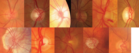
Clinicians who rely on the ISNT rule when assessing the optic nerve—for signs of early glaucoma, for example—should remember it doesn’t necessarily apply to all patients. While the rule states optic nerves typically show a larger rim width inferior, superior, nasal and then temporal, a new study highlights just how often patients deviate from this: within this particular study population, only 37.0% of rim assessments and 43.8% of retinal nerve fiber layer (RNFL) measurements follow the rule, according to the researchers.
 |
| Optic nerves come in all shapes and sizes, making the ISNT rule tough to follow. Can you tell which are normal, anomalous but non-glaucomatous and glaucomatous? Click image to enlarge. Photos: Jarett Mazzarella, OD |
“As we know, there is a wide variance of normal nerve anatomy, which makes diagnosing early glaucoma difficult in some patients,” says Jarett Mazzarella, OD, who practices in the VA Health Care System in Salisbury, NC. “Although a number of patients in the study did not conform to the standard rule, in my opinion it does not invalidate the ISNT rule since the areas of the rim we are concerned with in early glaucoma are the inferior or superior rim.”
Researchers looked at 110 normal subjects and found a larger or smaller nasal sector was one of the most significant causes of deviation, with 10.9% of subjects having a wider nasal rim than inferior, 29.4% with a wider nasal rim than superior, 14.7% with a narrower nasal rim than temporal and 42.9% having thinner nasal RNFLs compared with the temporal quadrant.
“We know glaucoma patients tend to lose neuroretinal rim on the superior and inferior rims, and in this study, excluding the nasal rim to modify the rule to the IST or IS made it apply to roughly 70%,” says Justin Cole, OD, of the VA Health Care System in Salisbury, NC. “So the rule still applies, but I also think we have really been using it as the ‘IS’ rule all along.”
For clinicians assessing patients for early signs of glaucoma, Dr. Mazzarella says the findings serve as a stark reminder of the importance of obtaining baseline readings, following for change over time and “not getting stuck in the mindset of always following traditional rules.”
“Early disease is the confounding factor between identifying abnormal structure vs. a normal variant. For example, we see this often with our OCT technology when a patient flags as abnormal on OCT RNFL or ganglion cell compared with the normative data values,” Dr. Mazzarella says. “Many of these patients never change or progress, which usually indicates a variant of normal anatomy. It comes down to establishing a baseline for that individual patient and watching for any indication of structural or functional progression over time, especially for the normal nerve that does not look so ‘typical.’”
“Each clinician must use their own judgment in identifying normal from abnormal cup-to-disc ratios and appearances, while also having the wherewithal to know that anatomical differences are common,” concludes Dr. Cole.
| Poon LYC, Valle DSD, Turalba AV, et al. The ISNT rule: how often does it apply to disc photographs and retinal nerve fiber layer measurements in the normal population? Am J Ophthalmol. 2017;184:19-27. |
Hot Tea’s Impact on GlaucomaTea drinkers have one more reason to brew another pot this winter. A new study found drinking hot, caffeinated tea may be associated with a lower risk of glaucoma. Data from the 2005-2006 National Health and Nutrition Examination Survey indicates hot tea-drinkers were 74% less likely to have glaucoma. The same was not true for coffee (caffeinated or decaffeinated) decaffeinated tea, iced tea or soft drinks. While the survey had a small number of patients diagnosed with glaucoma and didn’t take into account other factors such as cup size, tea type or brewing time, the researchers speculate the tea’s antioxidants and anti-inflammatory and neuroprotective chemicals may play a role. Wu CM, Wu AM, Tseng VL, et al. Frequency of a diagnosis of glaucoma in individuals who consume coffee, tea and/or soft drinks. British J Ophthalmol. December 14, 2017. [Epub ahead of print]. |
Excessive Exercise May Raise Men’s AMD Risk
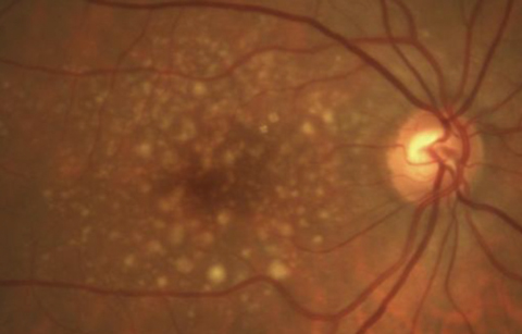
A new study suggests exercising five or more times a week may increase a man’s risk of neovascular age-related macular degeneration (AMD). Researchers in Japan looked at 211,960 patients ages 45 to 79 at baseline and then seven to 10 years later and found men ages 45 to 64 who exercised vigorously had a 54% increased risk of neovascular AMD compared with men in the non-exercise group. They did not find the same association in women. These results come after adjusting for factors such as age, medical history and body mass index.  |
| Vigorous exercise may increase a man’s risk of neovascular AMD, as seen here. Photo: Andrew Rixon, OD |
While neither previous research nor this study provide further insight into the possible reasons behind the association, the investigators speculate excessive exercise may affect the patient’s choroid. Still, no strong biological rationale exists for this finding yet.
“However, the authors were cautious about the results and recommended additional studies,” says Sherrol Reynolds, OD, an associate professor of optometry at Nova Southeastern University College of Optometry. “Before clinical recommendations are made about limiting physical activity or discussing caution with rigorous activity and neovascular AMD, more research is necessary.”
| Rim TH, Kim HK, Kim JW, et al. A nationwide cohort study on the association between past physical activity and neovascular age-related macular degeneration in an East Asian population. JAMA Ophthalmol. December 14, 2017. [Epub]. |
Macular Damage Mechanism Discovered
Researchers at the University of Virginia School of Medicine have uncovered one of the first triggers of inflammation in macular degeneration, a common enzyme called cyclic GMP-AMP synthase (cGAS).  |
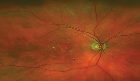
| Patient with AMD may one day have a new treatment option to combat geographic atrophy, as seen here. Photo: Sara Weidmayer, OD |
“This research found that, in human cell cultures and mice in vivo, certain proteins including the DNA sensing enzyme cGAS, played a role in activating an inflammatory immune response that ultimately leads to retinal pigmented epithelium (RPE) cell death,” says Sara Weidmayer, OD, who works at the VA Ann Arbor Healthcare System in Ann Arbor, Mich., and is a clinical assistant professor at the Kellogg Eye Center, Department of Ophthalmology and Visual Sciences at the University of Michigan.
“These proteins were found in higher levels in patients with geographic atrophy (GA), so the findings suggest that they are involved in the pathogenesis of GA.”
While cGAS is well known for its role in detecting foreign DNA and initiating the immune response to infections, researchers were surprised to discover its impact on dry age-related macular degeneration. Without any foreign invasion to activate cGAS, elevated levels in the RPE of eyes with GA suggests the enzyme may play a bigger part in the body’s response to noninfectious human disease.
“These findings continue to provide insight into the understanding of the mechanisms of cell damage and death in patients with geographic atrophy,” says Dr. Weidmayer. “Watching so many patients progressively lose their vision due to GA, with very little to offer in the way of treatment, is disheartening; the prospect of someday being able to employ a treatment to block an enzyme which drives the disease is certainly exciting for the entire eye care community.”
“This research may prompt the development of medications to block or inhibit cGAS, which could ultimately change the trajectory of visual outcomes for GA patients,” Dr. Weidmayer adds. “Hopefully this research will serve as a step on the path to the goal of individualized, precision medicine.”
Kerur N, Fukuda S, Banerjee D, et al. cGAS drives noncanonical-inflammasome activation in age-related macular degeneration. Nature Medicine. November 27, 2017. [Epub]. |
In the newsThe FDA recently approved Luxturna (voretigene neparvovec-rzyl, Spark Therapeutics), a directly administered gene therapy that targets biallelic RPE65 mutation-associated retinal dystrophy. The therapy is designed to deliver a normal copy of the gene to retinal cells to restore vision loss. While the approval provides hope for patients, the $425,000 per eye price tag stands as a significant hurdle. Scutti S. Gene therapy for rare retinal disorder to cost $425,000 per eye. CNN. www.cnn.com/2018/01/03/health/luxturna-price-blindness-drug-bn/index.html. Accessed January 4, 2018. A study 167 patients with suspected bacterial endophthalmitis after cataract surgery found intravitreal dexamethasone provided no improved visual acuity. Patients were treated twice with intravitreal injections of 0.2mg vancomycin and 0.05mg gentamicin, followed by either 400µg dexamethasone sodium diphosphate or placebo. The four-week, 10-week, six-month and 12-month follow ups showed no significant difference in best-corrected visual acuity. Manning S, Ugahary LC, Lindstedt EW, et al. A prospective multicentre randomized placebo-controlled superiority trial in patients with suspected bacterial endophthalmitis after cataract surgery on the adjuvant use of intravitreal dexamethasone to intravitreal antibiotics. Acta Ophthalmol. December 7, 2017. [Epub]. New research suggests a desktop humidifier may help patients with dry eye symptoms during continuous computer use. Investigators measured noninvasive tear break-up time (NTBUT) in patients who did and did not use a desktop humidifier for an hour of computer use and found improved NTBUT in the humidifier users compared with those without a humidifier. Wang MT, Chan E, Ea L, et al. Randomized trial of desktop humidifier for dry eye relief in computer users. Optom Vis Sci. 2017;94(11):1052-7. |


