 |
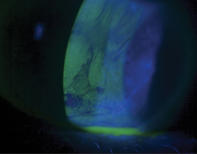
This study showed a correlation between vitamin D level and tear parameters in children with type 1 diabetes. Photo: Jack Schaeffer, OD. Click image to enlarge. |
Tear function parameter changes in type 1 diabetes have been previously investigated, but very few studies have looked into changes to the ocular surface in such patients. In a recent study, researchers evaluated dry eye test parameters of affected kids with and without vitamin D deficiency.
A total of 90 eyes of 90 pediatric patients with type 1 diabetes were compared with 80 eyes of 80 controls. Ocular Surface Disease Index (OSDI), Schirmer test, tear film breakup time (TFBUT), corneal staining score and anterior segment optical coherence tomography were used to determine the dry eye test parameters. The study group was divided into two subgroups based on vitamin D status: those with vitamin D deficiency and those with normal vitamin D levels.
Lower OSDI scores, TFBUT, Schirmer test scores and tear meniscus parameters were found in pediatric type 1 diabetes patients without dry eye symptoms. Additionally, the tear meniscus dimensions decreased significantly in the diabetes patients with vitamin deficiency compared with the controls and those without.
A previous study similarly reported a decrease in the TFBUT and Schirmer score in children with type 1 diabetes and found this to be correlated with the duration of diabetes.
Other studies observed vitamin D receptors in the corneal epithelium, the endothelium and the retinal pigmentary epithelium, making it possible that vitamin D may regulate the tear secretion of the lacrimal glands through similar types of mechanisms, the authors suggested. “However, the association that exists between dry eye syndrome and vitamin D is not yet understood,” they explained. “Except for the OSDI scoring, the other tests were significantly in favor of dry eye when compared with the DM patient group with normal vitamin D levels.”
“The correlation between the vitamin D level and the tear parameters showed that physicians must be more sensitive to this issue,” the authors concluded. “Therefore, in addition to the use of OSDI scores, TFBUT and corneal staining assessment, measurement of size of the tear meniscus by the use of objective and noninvasive Spectralis OCT for screening and diagnosis should be considered.”
Aydemir GA, Aydemir E, Asik A. Changes in tear meniscus analysis of children who have type 1 diabetes mellitus, with and without vitamin D deficiency. Cornea. November 23, 2021. [Epub ahead of print]. |

