History
A 24-year-old white male contact lens wearer presented for a routine eye examination. He had no visual complaints but was concerned about chronic redness in both eyes, which he said had been a recurrent problem during the last year.
His extended ocular history did not offer any obvious etiology. His systemic history was unremarkable and noncontributory. He denied using any medication.
Diagnostic Data
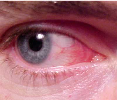
His best-corrected contact lens acuity was 20/20 O.U. at distance and near. The photograph demonstrates the noticeable external findings. There was no evidence of afferent pupillary defect. Anterior segment examination uncovered a mild papillary reaction of the superior palpebral conjunctivae, but there was no significant corneal staining and no infiltration. Intraocular pressures measured 18mm Hg O.U. The dilated fundus examination was normal.
|
|
| The patients anterior segment exam uncovered a mild papillary reaction of the superior palpebral conjunctivae, but there was no significant corneal staining and no infiltration. |
Your Diagnosis
How would you approach this case? Does this patient require additional tests?
What is your diagnosis? How would you manage this patient? What is the likely prognosis?
Thanks to Jason Hales, O.D., United States Army, for contributing this case.
Discussion
Additional testing included revisiting the patients history to better understand the potential relationship of the contact lenses and/or the contact lens solutions to the signs and symptoms. We also palpated the lymph nodes to rule out viral disease, observed the tear prism to rule out dry eye and used Schirmer test strips to demonstrate normal wetting O.U.
The diagnosis in this case, virtually by exclusion, is allergic conjunctivitis. We initiated a step-based action plan to eliminate the signs and symptoms with the least amount of therapy.
First, we instructed the patient discontinue contact lens wear and apply cold compresses q.i.d. along with artificial tears to soothe, dilute and mechanically wash the ocular surface. This proved to be only intermittently effective.
Next, we added a topical mast cell stabilizer/antihistamine combination, Patanol (olopatadine, Alcon) b.i.d. O.U. While this stopped most of the symptoms, it did not eliminate the injection.
Finally, we prescribed a mild topical steroid, Lotemax (loteprednol, Bausch & Lomb), q.i.d. O.U. This resolved the condition entirely. We slowly tapered the steroid over the course of one month. We refit the patient in contact lenses (CSI, CIBA Vision) and changed his solution to a hypoallergenic one (Clear Care, CIBA Vision). We advised the patient to maintain good contact lens habits and hygiene and to lubricate his eyes with artificial tears (Cellufresh, Allergan) frequently as needed throughout the day, with a minimum of q.i.d. It has been six months since the initial episode, and there have been no recurrences.
The human allergic response has varied objective signs and physical symptoms. Ocular allergic conditions vary from the subtle signs of itchy, watery eyes with mild hyperemia to extensive inflammatory interactions between the ocular coats and adnexa. Patient symptomatology often includes itching, burning and tearing of the eyes with watery discharge. In most cases, a history of allergies (seasonal or other) can be elicited.1-4 The important observable clinical signs include tissue swelling (chemosis); red, edematous eyelids; conjunctival papillae; and a lack of palpable preauricular node.
The allergic response is classically considered to be an over-reaction of the bodys immune system to foreign substances known as immunogens or allergens.1-3 The response can be innate or acquired.
The mast cell is the key component to the ocular allergic response. When mast cells interact with specific allergens, they open like a lock being opened by a key (degranulation), and discharge chemical substances called mediators into the surrounding tissues.4 The primary chemical mediators include:1-4
Histamine, which is responsible for increased vascular permeability, vasodilation, bronchial contraction and increased secretion of mucus.
Neutral proteases, which generate other inflammatory mediators.
Arachadonic acid, a crucial component of the cyclooxygenase pathway.
Because there are many strata of ocular allergic reactions, management is primarily aimed at reducing symptomatology. Perhaps the easiest and most effective treatment for allergic conjunctivitis is elimination or avoidance of the potentially offending allergen. Cold compress and artificial tears (drops and ointments) soothe and lubricate on an as-needed basis. Medical therapy includes:1-4
Topical decongestants. These produce vasconstriction and reduce hyperemia, chemosis and other symptoms by retarding the release of the chemical mediators into the tissues from the bloodstream. Examples: naphazoline and phenylephrine, b.i.d. to q.i.d. Given the armamentarium of ophthalmic medications, these agents are less frequently used.
Antihistamines. Topical and oral antihistamines are also excellent therapies. Examples: topicalAlamast (pemirolast, Vistakon), Alocril (nedocromil, Allergan) and Emadine (emedastine, Alcon), b.i.d. to q.i.d.; oralBenadryl (diphenhydramine, Pfizer) 25mg p.o. t.i.d. and Claritin (loratadine, Schering-Plough) 10mg p.o. q.d.
Topical antihistamine/mast cell stabilizers. The antihistamine addresses the acute symptoms, and the mast cell stabilizer prevents subsequent allergy expression. Examples: Elestat (epinastine, Allergan), Optivar (azelastine, MedPointe), Patanol (olopatadine, Alcon) and Zaditor (ketotifen, Novartis), b.i.d. to q.i.d.
Topical nonsteroidal anti-inflammatory drugs. NSAIDs may offer relief in moderate cases. Examples: Acular LS (ketorolac, Allergan) and Voltaren (diclofenac, Novartis), b.i.d. to q.i.d.
Topical steroidal preparations. Topical steroids are reserved for the most severe presentations. Examples: Alrex (loteprednol 0.2%, Bausch & Lomb), Flarex (fluorometholone acetate, Alcon), FML (fluorometholone alcohol, Allergan), Lotemax (loteprednol 0.5%, Bausch & Lomb), Inflamase Forte (prednisolone sodium, Novartis), Pred Mild (prednisolone acetate 0.12%, Allergan), Pred Forte (prednisolone acetate 1%, Allergan) and Vexol (rimexolone, Alcon), b.i.d. to q.i.d.
Educate patients who have a history of seasonal allergic conjunctivitis to avoid the substances that precipitate symptoms. In severe cases, these patients may be treated four weeks in advance with loading doses of a topical mast cell stabilizer or mast cell stabilizer/antihistamine combination q.i.d. O.U. to retard the degranulation process and to slow, reduce or eliminate the debilitating correlates. Patients placed on supportive therapies may be followed as needed. Follow patients taking topical NSAIDs one to two weeks after the start of therapy.
1. Bonini S. Allergic conjunctivitis: the forgotten disease. Chem Immunol Allergy 2006;91:110-20.
2. Butrus S, Portela R. Ocular allergy: diagnosis and treatment. Ophthalmol Clin North Am 2005 Dec;18(4):485-92, v. Review.
3. Bielory L. Allergic diseases of the eye. Med Clin North Am 2006 Jan;90(1):129-48. Review.
4. Leonardi A. Emerging drugs for ocular allergy. Expert Opin Emerg Drugs 2005 Aug;10(3):505-20.


