 History
A 27-year-old black male presented for an examination following an elbow to his right eye during a basketball game. His systemic and ocular histories were noncontributory. He denied taking medications and did not report any allergies.
Diagnostic Data
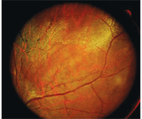
His best-corrected visual acuity was 20/40 O.D. and 20/20 O.S. at distance and near. External examination was normal, with no evidence of afferent pupillary defect. His anterior segment structures were normal. Intraocular pressure measured 15mm Hg O.U. The pertinent fundus findings are illustrated in the photograph.
Your Diagnosis

This 27-year-old male presented
after suffering an elbow to his right eye during a basketball game. Did
this personal foul cause substantial ocular damage?
How would you approach this case? Does this patient require any additional tests? What is your diagnosis? How would you manage this patient? What’s the likely prognosis?
Discussion
The diagnosis in this case is commotio retinae of the right eye. Additional testing might include close inspection of the orbit for emphysema or crepitus; X-ray or CT scan to rule out fracture; close inspection of anterior chamber for inflammation or hyphema; close inspection of the posterior peripheral fundus to rule out retinal breaks or tears; and photodocumentation. Gonioscopy is not advisable immediately after a patient suffers blunt trauma. Instead, gonioscopy should be performed when the eye has returned to homeostasis to assess for damage that might suggest the signs and symptoms of late-traumatic glaucoma. Keep in mind that patients who develop commotio retinae need to be monitored regularly for glaucoma for the rest of their lives.
Commotio retinae, or Berlin's edema, occurs as a result of a contrecoup injury. Relatively well-defined areas of retinal opacification are observable following an injury to the globe or its adbexa. Experimental evidence and clinical observation suggest the retinal changes are secondary to deep sensory retinal edema.1-5
Histologically, photoreceptor cell bodies (specifically the photoreceptor outer segments and their connections to the outer plexiform layer) are damaged early in the process.3-5 As the condition resolves, the retinal pigment epithelium phagocytizes the damaged outer segment material.4,5 This permits the retinal pigment epithelium to undergo hyperplasia, with the potential for migration into the neurosensory retina depending on the severity of the initial trauma.3,5
The process may resolve completely without sequelae.1-3 In unfortunate circumstances, however, injury to the photoreceptors may cause permanent visual loss.1-5 Cystoid macular degeneration, with cyst and hole formation, may occur months or years later.1,2,5 Additionally, microcysts (small accumulations of intraretinal fluid) may initiate a tissue breakdown, resulting in variations of macular pathology.1-5
We treated the patient with in-office cycloplegia and mild topical steroidal medication q.i.d. We recommended cold compresses and rest q.i.d. in addition to non-antiplatelet analgesics, as necessary, to mitigate discomfort.
We performed photodocumentation as well as educated the patient about potential complications and the usual course of resolution. We dispensed an Amsler grid for home monitoring and scheduled four follow-up visits during the next two weeks.
At the fourth follow-up, we found no complications and noted complete resolution, with a full restoration of pre-injury visual acuity.
1. Aldave AJ, Gertner GS, Davis GH, et al. Bungee cord-associated ocular trauma. Ophthalmology. 2001 Apr;108(4):788-92.2. Williams DF, Mieler WF, Williams GA. Posterior segment manifestations of ocular trauma. Retina. 1990;10 Suppl 1:S35-44.
3. Sony P, Venkatesh P, Gadaginamath S, et al. Optical coherence tomography findings in commotio retina. Clin Experiment Ophthalmol. 2006 Aug;34(6):621-3.
4. Meyer CH, Rodrigues EB, Mennel S. Acute commotio retinae determined by cross-sectional optical coherence tomography. Eur J Ophthalmol. 2003 Nov-Dec;13(9-10):816-8.
5. Carpineto P, Ciancaglini M, Aharrh-Gnama A, et al. Optical coherence tomography and fundus microperimetry imaging of spontaneous closure of traumatic macular hole: a case report. Eur J Ophthalmol. 2005 Jan-Feb;15(1):165-9.

