 |
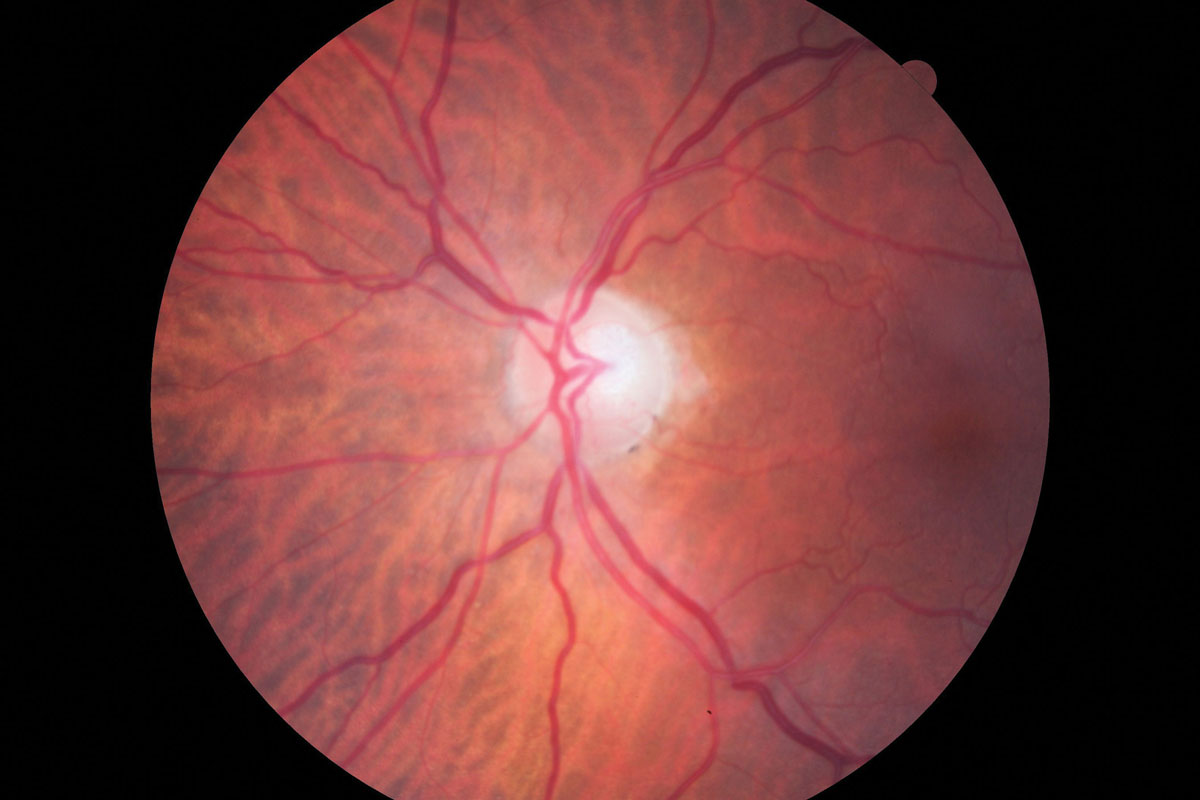
Peripapillary vessel density aids in predicting visual function. Click image to enlarge. |
Few studies have evaluated the clinical implications of postoperative microvascular changes in association with the visual prognosis in glaucoma patients. The aim of this study was to determine the factors associated with visual field (VF) deterioration after trabeculectomy, including the peripapillary vessel density (pVD) and macular vessel density (mVD) changes assessed by optical coherence tomography angiography (OCT-A).
Primary open-angle glaucoma (POAG) patients with more than two years of follow-up after trabeculectomy were included. pVD was calculated in a region defined as a 750μm-wide elliptical annulus extending from the optic disc boundary. mVD was calculated in the parafoveal (1-3mm) and perifoveal (3-6mm) regions. VF deterioration was defined as the rate of mean deviation (MD) worse than -1.5dB/year. The change rates of pVD and mVD were compared between the deteriorated VF and non-deteriorated VF groups.
VF deterioration was noted in 14 of the 65 eyes that underwent trabeculectomy. The pVD reduction rate was significantly greater in the deteriorated VF group than in the non-deteriorated VF group, while that of parafoveal and perifoveal VD did not show a significant difference. Linear regression analysis showed that the postoperative MD reduction rate was significantly associated with the rate of pVD reduction, while other clinical parameters and preoperative vascular parameters did not show any association. Eyes with greater loss of peripapillary retinal circulation after trabeculectomy tended to exhibit VF deterioration.
“Our study showed that the assessment of peripapillary retinal circulation can be used as a predictor of visual function after IOP-lowering surgery,” the authors concluded. “Considering that most glaucoma patients who have undergone trabeculectomy already have a substantial loss of VF, a biomarker reflecting visual function change is important in the care of advanced glaucoma patients.”
The authors suggested the use of OCT-A-measured pVD as the biomarker for predicting visual function after trabeculectomy.
Yoon J, Sung KR, Shin JW. Changes in peripapillary and macular vessel densities and their relationship with visual field progression after trabeculectomy. J Clin Med. 2021;10(24):5862. |

