 |
|
Scientists at the University of Iowa
recently found a unique macular dystrophy with intraretinal pigment
spots, cysts and hemorrhages.
|
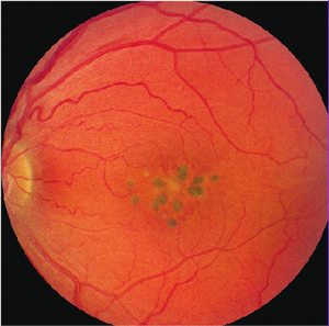
The researchers uncovered a peculiar macular dystrophy with intraretinal pigment spots, cysts and hemorrhages in a 24-year-old female and decided to examine her family members for similar presentations. The researchers performed dilated fundus examinations, optical coherence tomography scans, fluorescein angiography and electroretinography on 17 of the patient’s relatives.
Upon testing, seven family members exhibited multiple central macular cystic spaces and densely pigmented spots within their retinas. Additionally, two relatives had active macular neovascularization and leakage.
Most importantly, however, the researchers confirmed that all findings were consistent with an autosomal dominant pattern of inheritance.
“Through our paper and by sharing pictures of what the affected eye looks like, we hope to find more people affected,” says lead author Vinit Mahajan, M.D., Ph.D., assistant professor of ophthalmology and visual sciences at the University of Iowa Roy J. and Lucille A. Carver College of Medicine.
“We also will work to find the gene that causes the condition, because this information could be very useful in eventually preventing or treating this and other diseases that affect the macula,” Dr. Mahajan says.
Mahajan VB, Russell SR, Stone EM. A new macular dystrophy with anomalous vascular development, pigment spots, cystic spaces, and neovascularization. Arch Ophthalmol. 2009 Nov;127(11):1449-57.

