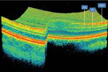
New research suggests that retinal thinning, as measured by OCT, can indicate how fast multiple sclerosis (MS) progresses—especially in the early course of the disease. Also, any eye doctor can measure it using commercially-available OCT equipment with retinal layer segmentation software.

OCT reveals thinning of the ganglion cell/inner plexiform layer, which indicates progression of multiple sclerosis.
In this study, conducted at the Johns Hopkins MS Center in Baltimore, 164 patients with MS and 59 healthy controls underwent spectral-domain OCT scans every six months, for an average of 21 months. Participants were also given MRI brain scans at baseline and at the yearly follow-up.
The researchers found that people with MS relapses had 42% faster thinning of the ganglion cell/inner plexiform (GCIP) layer than people with MS who had no relapses. Also, people with MS who had inflammatory gadolinium-enhancing lesions experienced 54% faster thinning, and those with new T2 lesions had 36% faster thinning than MS patients without these conditions.
People whose level of disability worsened during the study experienced 37% more thinning than those who had no changes in their level of disability. Further, those who had the disease less than five years showed 43% faster thinning than individuals who had the disease more than five years.
Looking to the future, the researchers concluded that OCT-derived GCIP thickness assessment could be used as an outcome measure for evaluating neuroprotective agents, particularly in early, active MS. “As more therapies are developed to slow the progression of MS, testing retinal thinning in the eyes may be helpful in evaluating how effective those therapies are,” says lead author Peter Calabresi, MD, director of the Johns Hopkins MS Center.
Ratchford JN, Saidha S, Sotirchos ES, et al. Active MS is associated with accelerated retinal ganglion cell/inner plexiform layer thinning. Neurology. 2013 Jan 1;80(1):47-54.

