A 66-year-old white female with a longstanding history of glaucoma presented for visual field testing, optic nerve scans, intraocular pressure evaluations and pachymetry measurements before undergoing a scheduled selective laser trabeculoplasty (SLT). She exhibited signs of progressive field loss and potential neuroretinal rim changes in her right eye, despite receiving topical glaucoma therapy.

Diagnostic Data
She was initially seen in 1998 as a new patient. She had complained of blurred vision in both eyes. Her medications included hydrochlorothiazide (HCTZ) and potassium supplementation, and she reported no known allergies. At that time, her best-corrected visual acuity measured 20/20 O.D., O.S. and O.U. through myopic astigmatic correction. Her pupils were equally round and reactive to light and accomodation, with no afferent defect. Her extraocular muscles were full in all positions of gaze.
A slit lamp examination of her anterior segments was essentially unremarkable. Her anterior chamber angles measured grade 4 open O.U., using Van Herick’s method. Her intraocular pressure was 27mm Hg O.D. and 25mm Hg O.S. at 10:30 a.m. Through dilated pupils, her crystalline lenses were clear O.U. Her cup-to-disc ratio was estimated to be 0.50 x 0.75 O.D. and 0.55 x 0.65 O.S. I noted thinning of the inferotemporal neuroretinal rims (O.D.>O.S.). The vasculature was characterized by mild
hypertensive retinopathy in both eyes. Her macular and peripheral retinal evaluations were normal O.U. Given her elevated IOP and suspect optic nerve appearances, I asked her to return for a full glaucoma evaluation.
At the glaucoma evaluation, her IOP was 26mm Hg O.D. and 23mm Hg O.S. Threshold visual fields were unreliable, with high fixation losses and high false positives. There were scattered paracentral areas of field depression, but given the poor reliability indices, we determined that the fields did not provide useful information. We also took stereoscopic nerve images. Gonioscopy demonstrated IV+ open angles O.U., with minimal trabecular pigmentation.
Initially, I prescribed 1 drop Betagan 0.5% (levobunolol, Allergan) O.U. q.d., because I believed that she clearly had glaucoma. Following treatment, her IOP averaged 16mm Hg O.D. and 15mm Hg O.S.
Over the years, she attended all scheduled follow-ups, which often included IOP measurement, threshold visual field studies, and eventually, conofocal scanning laser ophthalmoscopy of her optic nerves. Pachymetry of her central corneal thickness measured 525µm O.D. and 518µm O.S. Also, Heidelberg Retina Tomography-3 (HRT-3, Heidelberg Engineering) scans confirmed the loss of the inferotemporal neuroretinal rims in both eyes.
Due to gradually and consistently elevated IOP, I eventually added 1 drop Travatan (travoprost, Alcon) O.U. h.s. to her topical beta blocker (which, at that time, was generic 0.5% timolol gel forming solution) in 2007. This reduced her IOP to an average of 14mm Hg O.U. for nearly 18 months. Visual fields, though variable and often less than reliable, and HRT-3 nerve scans remained stable during this time.
In 2009, her IOP crept back into the low 20s O.U., and I instructed her to continue dosing the 1 drop Travatan (now Travatan-Z) O.U. h.s., discontinue the 0.5% timolol, and begin instilling 1 drop Cosopt (dorzolamide and timolol, Merck) O.U. b.i.d. She tolerated the medication change well, and her post-treatment IOP was again reduced to the mid teens O.U.
Then, at a scheduled follow-up in the summer of 2010, her IOP measured 24mm Hg O.D. and 20mm Hg O.S. Gonioscopy was similar to that of 1998, except for a slight increase in the amount of trabecular pigmentation. HRT-3 scans were stable and––unsurprisingly––visual fields were not very reliable. While this patient had documented
episodes of increased IOP in the past, I was not convinced that her IOP was consistently elevated at this time, because this was the first visit where her IOP was elevated since we adjusted her regimen in 2009.

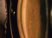
Like this individual, our patient had an open angle and minimal trabecular
pigmentation O.D. on gonioscopy.
Giving the situation the benefit of the doubt, I did not change her therapy right away. I re-stressed the importance of compliance, and explained that if her IOP remained elevated at the next visit, she would require an SLT procedure in her right eye. We scheduled her for a follow-up in four weeks.
Six weeks later, she presented for the follow-up with an IOP of 25mm Hg O.D. and 21mm Hg O.S. The HRT-3 scan and topographical change analysis revealed a slight decrease in inferotemporal neuroretinal rim volume in her right eye and a stable neuroretinal rim in her left eye. Again, visual fields were unclear, with low reliability indices. At this point, we scheduled the patient for an SLT procedure in her right eye.
I told the patient that she would be seeing the surgeon for one visit, and said that the SLT procedure was painless and would be performed in the office. The patient was scheduled for a follow-up visit in our office two weeks after the surgery.
Discussion
This is a pretty straightforward case that highlights the nature of glaucoma therapy––even with constant vigilance and topical therapy, some patients tend to progress. Having a good “plan B” in place is essential for these patients. And, at least in the U.S., plan B often includes SLT or argon laser trabeculoplasty (ALT).
How you participate in the postoperative laser care of your glaucoma patients as well as how involved you want to be in the care process varies from clinician to clinician. Some O.D.s are more comfortable referring the patient to the surgeon with the caveat of having the patient return for follow-up after the laser procedure has been performed and a sustained IOP reduction has been achieved. On the other hand, there are O.D.s, like myself, who want to maintain a very hands-on approach to patient care––both before and, particularly, immediately after the laser therapy. Ideally, I would like to limit our patient’s experiences with the surgeon to only the one office visit for the actual laser procedure.
The postoperative course of both SLT and ALT patients is very similar. Postoperative inflammation, though unlikely, can occur. In general, ALT will cause a faster IOP reduction than SLT. With ALT, the effects from the laser can be seen in two to four weeks, whereas the full benefit from SLT may not be achieved for six to eight weeks. Either way, the patient needs to be seen regularly during the next one to three months to determine the therapeutic effect of the laser. And certainly, optometrists are more than capable of determining that therapeutic benefit.
Always be sure to provide the laser surgeon with a comprehensive history to reduce the number of visits the patient must make to his or her office. Include copies of all visual fields tests, optic nerve scans and glaucoma flow sheets so that the surgeon can become comfortable with your assessment of the patient and more clearly see that the patient does, in fact, need laser therapy. Without that information, the laser surgeon could be hard pressed to defend his or her position on performing laser surgery.
By the time our patient presented for the SLT, the surgeon had already received our copies of her visual fields, optic nerve scans and glaucoma flow sheets. We scheduled her for a two-week follow-up after the visit with the surgeon. But, as often happens in busy practices, the patient did not return to our office for nearly six weeks.
When I asked her about the delay in returning to the office, she told me that the laser surgeon had seen her for two pre-laser visits and one post-laser visit. Oddly enough, I was unaware of this. Furthermore, I learned that prior to initiating the laser therapy, the surgeon repeated visual fields, OCT nerve fiber layer scans, gonioscopy and two pressure readings.
And, following the laser procedure, the patient was also seen for two IOP readings.
This was not how I expected things to go. I intended for the surgeon to see the patient just once to perform the actual laser procedure, or maybe once more after the procedure if any complications arose. There is no reason, in my opinion, why visual fields, nerve scans and multiple IOP checks need to be done again, especially for a patient with a long, documented history of visits and trends over 12 years. Needless to say, the surgeon and I had a chat that day.
It is appropriate and within reason to voice any concerns you might have with a surgeon regarding perioperative patient care. Always remain cognizant of the surgeon’s concerns; however, if you cannot come to terms about sharing care in a manner that suits you, it may be beneficial to seek the services of another surgeon who does employ a similar approach to patient care.

