You know the situation: It’s Friday afternoon with one last exam to do, and you’re home free. And then you notice the last patient’s chief complaint: dry eye. Does this complaint fill you with dread or make you wish for a four-day work week? Or, does it excite you about the many future appointments you and this patient will share?
If you’re like most eye care professionals, you recognize dry eye disease as a challenging problem, not easily fixed with over-the-counter tears at your local big box store. Dry eye disease is also extremely common in our profession. By one estimate, 47% of patients visiting an optometrist’s office have complaints of dry eye.1
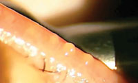 | |
| Because the majority of patients with dry eye have meibomian gland dysfunction, most dry eye patients can benefit from meibomian gland expression. |
One of the more prevalent forms of dry eye disease is meibomian gland dysfunction (MGD).2 In fact, dry eye and MGD are viewed by many experts as essentially synonymous. Although warm compresses, lipid-based artificial tears and oral doxycycline, among others, can help, you should also consider meibomian gland (MG) expression as a critical part of your regimen when diagnosing and treating this disease.
Fortunately, expression of the meibomian glands is one of the easiest procedures to perform and can produce lasting, noticeable results. This article—the fifth article in a six-part, print-and-video, instructional series—will show you how it’s done.
Understand the Gland
Before you grab your cotton-tip applicators or Mastrota paddle (Ocusoft), let’s discuss the causes of MGD.
The recently-completed International Workshop on Meibomian Gland Dysfunction identified hyperkeratinization of the meibomian gland duct as a primary cause of MGD.3 They found that bacteria located on the lid margin and within the glands produce enzymes that aggravate and incite keratinization of the gland epithelium. These enzymes also cause the lipids to be more viscous, which may lead to obstruction. Once the ducts are obstructed, the glands themselves may atrophy.3
Ophthalmic factors that may contribute to increased MGD include anterior blepharitis, Demodex infection and contact lens wear. Systemic factors, such as menopause or aging, and medications such as antihistamines, antidepressants, certain acne treatments and postmenopausal hormone therapy also have been shown to play a role in MGD.2
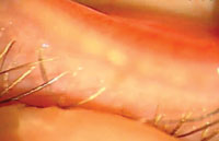 | |
| Meibomian glands that are capped with a white sebaceous material point toward the diagnosis of meibomian gland dysfunction. | |
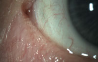 | |
| Another common sign of meibomian gland dysfunction is frothy or foamy tears in the tear lake or meniscus. |
On top of all these factors, the evolution of technology—including smartphones, tablets, computers and video games—may be increasing the prevalence of MGD. Studies show we blink less often and less fully when on the computer, which can lead to MGD.4
Who Needs Their Glands Expressed?
The first step is to identify which patients will most benefit from MG expression. In our opinion, nearly all patients who have dry eye symptoms or who are being worked up for dry eye disease could benefit from this easy procedure. Paul Karpecki, OD—who was instrumental in guiding us in setting up our dry eye clinic at the Oklahoma College of Optometry—recommends three items you’ll need if you want to get started in managing ocular surface disease and dry eye:
1. A good questionnaire
2. Vital dyes or stains
3. Meibomian gland expression
Most patients who present with MGD complain of dry or gritty eyes that are worse in the morning and may become less noticeable throughout the day.1 They may also complain of red, inflamed eyes. Fortunately, MGD is fairly easy to diagnose, often requiring only a quick slit lamp exam of the meibomian gland pores and instillation of fluorescein to determine the tear break-up time. Meibomian glands that are capped with a white sebaceous material, a cornea that exhibits a decreased tear break-up time, or both, are common signs pointing toward the diagnosis of MGD.
One of the other common signs of MGD is frothy or foamy tears in the tear lake or meniscus. In our clinical experience, there is nothing else that causes this foam other than MGD. While the exact mechanism that produces frothy tears is unknown, some experts believe that MGD causes the secretion of a foamy, surfactant-like material rather than healthy meibomian gland oil. Others hypothesize that the frothy presentation results from saponification, where bacterial enzymes react with tear lipids to form a soapy discharge. Regardless, both theories attribute the fundamental cause of frothy or foamy tears to MGD.
Other signs of MGD include increased tear film osmolarity, ocular surface staining, meibum that has a turbid or toothpaste-like consistency, and fluctuating visual acuity.2
Traditionally, MGD was treated with warm compresses, lid scrubs and artificial tears. But a recent report from the International Workshop on Meibomian Gland Dysfunction recommends performing meibomian gland expression at the earliest clinical signs of MGD.2 This critical step will help you understand the severity of the patient’s MGD and will guide you in your treatment approach.
Gland Anatomy and Meibum Appearance
Now that you’re convinced you should be performing MG expression on your patients, let’s talk about where to express the glands—and to do that, we need to review the anatomy.
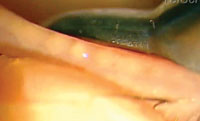 | |
| Healthy meibomian glands produce meibum that appears like olive oil or baby oil. But the cloudy meibum seen here is an early sign of MGD. |
The meibomian glands are sebaceous or oil-producing glands located in the tarsal plate of the eyelid. The gland lobes or acini are grouped together and are attached to a large central duct that terminates in a pore on the eyelid margin, posterior to the eyelid cilia. These glands receive parasympathetic innervation to make meibum and, under normal circumstances, eyelid contraction aids in secretion and distribution of the meibum across the surface of the eye.5 This simple fact helps to explain why today’s technology could be contributing to MGD because we not only blink less often but also less fully when viewing phones, computers or tablets.4
Meibum comprises the most anterior aspect of the tear film and stabilizes the tear film by preventing evaporation of the aqueous layer. Under healthy conditions, meibum consistency is similar to olive oil or baby oil in color and viscosity.
If it’s turbid or cloudy in color, that’s an early sign of MGD. If it becomes cheesy or toothpaste-like in texture, that is an indication of more advanced MGD. Severely plugged, non-functioning or atrophic glands are the most advanced signs of MGD. In these cases, pushing on the glands reveals no expression.3
Types of MG Expression
Meibomian gland expression can be divided into two groups: diagnostic and therapeutic.
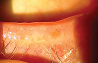 | |
| To express the inferior meibomian glands, pull the nasal aspect of the lower lid inferiorly to expose the meibomian gland pores and palpebral conjunctiva. | |
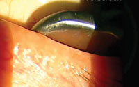 | |
| Apply the expressor paddle (or cotton-tip applicator) to the palpebral conjunctiva at a point midway between the fornix and the lashes. |
• Diagnostic expression is generally performed before therapeutic expression and is used to determine if the patient has MGD and, more specifically, to determine the stage of severity. It requires only a finger or cotton-tip applicator pressed and held against the inferior eyelid adnexa for approximately five to 15 seconds. In your busy practice, expressing lateral, central and medial aspects of all four eyelids may seem time-consuming and daunting. Fear not, as clinical experience shows it’s the central to nasal inferior lids that are the most important to express to get an idea of the severity of the patient’s MGD.2 A larger tool such as a Mastrota paddle also helps increase efficiency.
After a few seconds, you should see meibum excreting from the glands. Oil that is clear with little color indicates healthy meibomian glands. Thick or discolored meibum indicates MGD and proceeding with therapeutic expression is probably a viable treatment option. However, if no meibum is excreted and the pores are clear, the glands may have atrophied. In this case, therapeutic expression produces little, if any, results.
• Therapeutic expression is a more complete expression of the glands and can increase quality meibum production and decrease dry eye symptoms. While similar to diagnostic expression, therapeutic expression is more thorough and performed to promote healthy meibum production. The steps for proper therapeutic gland expression are outlined below.
Equipment
Meibomian gland expression requires few specialized tools. Positive results can be had with just a cotton-tip applicator and your finger. However, we recommend at least one Mastrota paddle or similar expressor paddle for more complete gland expression and enhanced patient comfort. Or, use two expressor paddles—one inside the lid and one outside the lid.
Besides the Mastrota paddle, other instruments are available for MG expression. The MG Expressor Kit (Gulden Ophthalmics) uses a flat paddle and a small roller to express the glands. The Arita MG Expressor (OcuSci), Collins Expressor Forceps (Bruder) and Maskin Meibum Expressor (Rhein Medical) are all types of forceps expressors that allow for one-handed use. The Thermal Pulsation System (Lipiflow) applies both heat and gentle pressure to express the meibomian glands. Finally, intense pulsed light (IPL) therapy, while not solely for meibomian gland expression, uses a specific wavelength of light to heat the blood vessels around the meibomian glands and liquefy the stagnant meibum. IPL is not new in the beauty/cosmetology industry, but recent ocular studies have found it to be effective for treating MGD and ocular rosacea.6
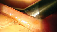 | |
| When you apply pressure to the inferior eyelid adnexa, meibum should soon emerge. However, if no meibum is excreted and the pores are clear, as shown here, the glands may have atrophied. | |
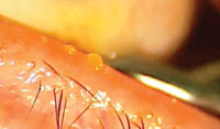 | |
| The thicker the meibum, the more advanced the MGD. This sludgy excretion indicates the meibomian glands are losing function. |
While magnification is necessary to diagnose MGD, you may find that performing gland expression outside of the slit lamp provides greater freedom of movement.
Set Up
Begin by instilling a drop of topical anesthetic, such as proparacaine, into both eyes. This is not usually needed for quick diagnostic expression, but it may be warranted, depending on the patient, for more extensive therapeutic expression.
Consider applying hot compresses for approximately 10 minutes prior to therapeutic expression. The heat softens or liquefies meibum, which improves the effectiveness of expression. Many types of heating masks are available, but we recommend the Bruder mask because it retains heat for 10 to 15 minutes.
To prevent corneal abrasions, have the patient look up for lower lid expression and down for upper lid expression.
How to Perform MG Expression
To express the inferior meibomian glands:
• Starting on the nasal aspect of the lower lid, pull the lid inferiorly to expose the meibomian gland pores and palpebral conjunctiva.
• Apply the expressor paddle or cotton-tip applicator (pre-moistened with saline) to the palpebral conjunctiva at a point midway between the fornix and the lashes.
• Release the lid and apply pressure to the inferior eyelid adnexa with a second cotton-tip applicator or expressor paddle.
• Roll the cotton-tip applicator or rock the paddle from the base of the meibomian glands to the pores on the eyelid margin.
• Pull the lower lid inferiorly and move the cotton-tip applicator or paddle to the next section of meibomian glands to be expressed.
• Follow the same maneuver for the remaining inferior meibomian glands.
To express the superior meibomian glands:
• With the patient looking down, lift the upper lid slightly off the globe.
• Insert the cotton-tip applicator or paddle under the lid, approximately midway between the superior fornix and lashes.
• Roll the cotton-tip applicator or rock the paddle inferiorly to express the glands.
• Repeat this process for the remaining superior glands.
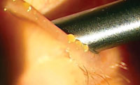 | |
| This thick meibum has a gooey, cheesy consistency, indicating more advanced MGD. | |
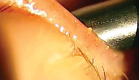 | |
| This very thick meibum was difficult to express and secreted from the glands with a dense, waxy consistency. |
After the Procedure
Consider instilling one to two drops of artificial tears per eye in the office immediately after expression. Prescribe your patient an artificial tear gel for use at home after MG expression. This provides some relief to the irritated palpebral conjunctiva.
After diagnostic or therapeutic meibomian gland expression has taken place in office, explain to the patient that long-term management of the MGD needs to be undertaken, as a one-time expression of the glands will not be a permanent cure. Strongly consider the following options in the long-term management of MGD:
1. Bruder mask or some type of warm compress mask that retains heat for 10 to 15 minutes, performed one to two times per day to continue evacuation of viscous meibum from the glands. In our clinical experience, patients note significant improvement with long-term use of these masks.
2. Recommend lipid-based tears such as Systane Balance (Alcon), Refresh Optive Advanced (Allergan), Retaine MGD (Ocusoft), or Soothe XP (Bausch + Lomb) to supplement the tear film and meibomian gland secretions.
3. Prescribe short-term steroid therapy in patients who exhibit signs and symptoms of posterior blepharitis (often caused by MGD).7 In our clinical experience, overnight use of Lotemax ointment (loteprednol 0.5%, Bausch + Lomb) or FML ointment (fluorometholone alcohol 0.1%, Allergan) has shown significant improvement in patient symptoms when used over the course of two to four weeks.
4. Prescribe tetracyclines to inhibit inflammation and stabilize the lipid layer of the tear film by decreasing free fatty acids. The International Workshop on Meibomian Gland Dysfunction recommends tetracyclines at all but the mildest stage of MGD.8 We typically use doxycycline 50mg BID for one to two months, then QD for one to two months. However, emerging research has shown that a short, five-day treatment of oral azithromycin may be more effective than doxycycline at treating MGD.9
5. Recommend an omega-3 fatty acid supplement to provide proper nutrition for meibum production. (Note: the American Heart Association recommends no more than 3g of omega fatty acids per day.)
Another supplement containing eicosapentaenoic acid (EPA), docosahexaeonic acid (DHA) and gamma linolenic acid (GLA) has been studied specifically for meibomian gland dysfunction and was shown to significantly improve signs and symptoms in MGD patients.10
Meibomian gland expression is both an easy and highly effective procedure for reducing symptoms and improving signs in MGD. Because MGD represents a significant portion of dry eye disease, proper management and treatment are essential for improving your patient’s ocular health.
With a little practice, you can put your patients on the road to producing viable meibum, increase their ocular comfort, and reduce your own dread of seeing another chief complaint of dry eyes.
Dr. Hatley is a clinical assistant professor at the Oklahoma College of Optometry and practices at the Cherokee Nation AMO Health Center in Salina, Okla.
Dr. Lighthizer is the assistant dean for clinical care services, director of continuing education, and chief of both the specialty care clinic and the electrodiagnostics clinic at the Oklahoma College of Optometry.
1. Fahmy A, Hauswirth SG, Hardten DR. Treatments for meibomian gland dysfunction. Ophth Management. 2012;16(9):25-6.2. Nichols KK, Foulks GN, Bron AJ, et al. The International Workshop on Meibomian Gland Dysfunction: Executive Summary. Invest Ophthalmol Vis Sci. 2011 Mar 30;52(4):1922-9.
3. Knop E, Knop N, Millar T, et al. The International Workshop on Meibomian Gland Dysfunction: Report of the subcommittee on anatomy, physiology, and pathophysiology of the meibomian gland. Invest Ophthalmol Vis Sci. 2011 Mar 30;52(4):1938-78.
4. Patel S, Henderson R, Bradley L, et al. Effect of visual display unit use on blink rate and tear stability. Optom Vis Sci. 1991 Nov;68(11):888-92.
5. Remington LA. Clinical Anatomy of the Visual System. 2nd ed. St. Louis, MO: Elsevier; 2005.
6. Toyos R, McGill W, Briscoe D. Intense pulsed light treatment for dry eye disease due to meibomian gland dysfunction; a 3-year retrospective study. Photomed Laser Surg. 2015 Jan;33(1):41-6.
7. Nelson JD, Shimazaki J, et al. The International Workshop on Meibomian Gland Dysfunction: Report of the Definition and Classificiation Subcommittee. Invest Ophthalmol Vis Sci. 2011 Mar 30;52(4):1930-37.
8. Geerling G, Tauber J, Baudouin C, et al. The International Workshop on Meibomian Gland Dysfunction: Report of the Subcommittee on Management and Treatment of Meibomian Gland Dysfunction. Invest Ophthalmol Vis Sci. 2011 Mar 30;52(4):2050-64
9. Kashkouli MB, Fazel AJ, Kiavash V, et al. Oral azithromycin versus doxycycline in meibomian gland dysfunction: a randomised double-masked open-label clinical trial. Br J Ophthalmol. 2015 Feb; 99(2):199-204.
10. Sheppard J, Pflugfelder S. Long-term supplementation with n-6 and n-3 PUFAs improves moderate-to-severe keratoconjunctivitis sicca: a randomized double-blind clinical trial. Cornea. 2013 Oct;32(10):1297-1304.

