 A 65-year-old Hispanic female presented for an annual eye exam. She complained of decreased vision in her right eye that had persisted for seven months. Her medical history was significant for arthritis and hypertension. She reported taking medications for these conditions, but could not remember the drugs’ names. When we last saw the patient 18 months earlier, her visual acuity measured 20/20 O.U.
At this examination, however, her best-corrected visual acuity was 20/50 O.D. and 20/20 O.S. Confrontation fields were full to careful finger counting O.U. Her pupils were equally round and reactive to light, with no afferent pupillary defect. Amsler grid testing showed central metamorphopsia in her right eye. The anterior segment examination was normal in both eyes.
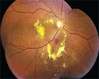
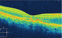
Dilated fundus exam showed moderate-sized cups, with good rim coloration and perfusion in both eyes. We noted obvious changes in her right eye (figure 1). We also performed a spectral domain optical coherence tomography scan of the right eye (figure 2). Her left eye was completely normal.
 |
 |
|
1, 2. Montage image of the right posterior pole shows extensive exudate in a circinate pattern (left). What is the underlying cause of the exudate? Spectral domain optical coherence tomography scan through the right macula (right). What does it reveal? | |
Take the Retina Quiz
1. Based on clinical appearance, what is the overall diagnosis?
a. Coats’ disease.
b. Macroarterial aneurysm.
c. Branch retinal vein occlusion (BRVO).
d. Neuroretinitis.
2. What does the OCT reveal?
a. Normal macula.
b. Mild cystoid macular edema (CME), increasing inferiorly.
c. Serous retinal detachment.
d. Ischemia.
3. How should this patient be managed?
a. Macular focal laser treatment.
b. Intravitreal Kenalog (triamcinolone acetonide, Bristol-Myers Squibb) injection.
c. Intravitreal Avastin (bevacizumab, Genentech) injection.
d. Observation.
4. What additional testing might provide the most useful information?
a. Visual field testing.
b. Fluorescein angiogram (FA).
c. Bartonella henselae blood testing.
d. Blood pressure testing.
For answers, see below.
Discussion
Our patient has extensive exudate in the macula and posterior pole of her right eye. The OCT of her fovea actually looks quite good, but she does have mild CME that becomes more evident on the inferior OCT rastar cuts.
So, why does she have this much exudate despite appearing completely normal 18 months earlier? If you look carefully along the inferior arcade, you can see a sheathed vein that extends inferiorly and begins where the artery crosses the vein. Based on this finding, we can conclude that our patient developed an inferotemporal BRVO several months earlier, which is now self-resolving.
If we had seen her when she first noticed the blurred vision seven months ago, there is no doubt that her retina would have looked quite different. Indeed, she would have exhibited the classic triangular or wedge-shaped hemorrhagic appearance extending peripherally where the artery crosses the vein.
Though the hemorrhaging has resolved, some leakage and incompetent retinal vessels are still spewing exudate throughout the posterior pole. The exudate has extended into the macula and is now causing CME.
So, how should our patient be managed? Based on results from the Branch Retinal Vein Occlusion Study, laser treatment is considered the standard of care for macular edema.1 The BRVO study showed that more than 50% of patients with macular edema improve to 20/40 or better with no treatment, but its results suggested that laser treatment should be considered for patients whose acuity does not improve significantly after a four to six month period.1
During the last few years, however, several new treatment options have challenged the traditional standard of care. Intravitreal Kenalog (IVK) garnered considerable attention in 2002 when new research suggested that patients with diabetes and recalcitrant macular edema experienced a substantial improvement in visual acuity and decreased retinal thickening after receiving treatment. With its success in treating diabetic macular edema, IVK emerged as a popular therapy for other conditions that cause macular edema, such as retinal vein occlusion. But, this new trend begged a major question––Is IVK better than laser for conditions that cause macular edema?
In an attempt to answer this question, the National Eye Institute launched the Standard Care vs. Corticosteroid for Retinal Vein Occlusion (SCORE) study. The SCORE study examined traditional standard of care (observation for CRVO, laser for BRVO) vs. 1mg or 4mg IVK injection for the treatment of macular edema secondary to either CRVO or BRVO in a randomized, controlled fashion.2,3 SCORE showed that patients in the CRVO group who were treated with IVK had a better visual outcome than simple observation.2 However, patients in the BRVO group who were treated with laser had a better visual outcome than those treated with IVK.3
At one-year follow-up of the BRVO group, the researchers documented a visual acuity improvement of three or more lines in 29% of patients who received laser treatment compared to 26% who received 1mg IVK and 27% who received 4mg IVK.3 At three-year follow-up, 20% to 30% of all patients in the BRVO group experienced a visual acuity gain of at least three lines. However, the researchers also noted more complications in patients who received IVK injection, including cataract development and increased IOP.3
Now that we have determined that laser is still a better treatment option than IVK for BRVO, what about the effects of anti-vascular endothelial growth factor (VEGF) agents?
Both Lucentis (ranibizumab, Genentech) and Avastin have shown great promise in the treatment of retinal vein occlusion because of their unique abilities to decrease capillary permeability. The Branch Retinal Vein Occlusion (BRAVO) study, for example, is a phase III, multicenter, randomized, controlled evaluation of the efficacy and safety of Lucentis vs. a sham injection in 397 patients with macular edema secondary to BRVO.4 At six-month follow-up, 55.2% of patients who received 0.3mg of Lucentis and 61.1% of patients who received 0.5mg of Lucentis gained 15 letters of best-corrected visual acuity, compared to just 28.8% of patients who received sham injections. Further studies are currently examining the treatment effect beyond a six-month period.
In this case, we sent the patient to a retinal specialist for treatment consideration. Approximately two weeks later, the patient received an intravitreal Avastin injection and was asked to return to our office for follow-up in one month. The surgeon also considered performing laser within the area of circinate exudate, but decided to wait and see the effect of the Avastin injection. He also believed that an FA would help establish the precise area of leakage as well as determine the presence of any ischemia within the macula. We plan to order an FA at the patient’s next follow-up.
1. The Branch Vein Occlusion Study Group. Argon laser photocoagulation for macular edema in branch vein occlusion. Am J Ophthalmol. 1984 Sep 15;98(3):271-82.
2. Ip MS, Scott IU, VanVeldhuisen PC, et al. A randomized trial comparing the efficacy and safety of intravitreal triamcinolone with observation to treat vision loss associated with macular edema secondary to central retinal vein occlusion: the Standard Care vs Corticosteroid for Retinal Vein Occlusion (SCORE) study report 5. Arch Ophthalmol. 2009 Sep;127(9):1101-14.
3. Scott IU, Ip MS, VanVeldhuisen PC, et al. A randomized trial comparing the efficacy and safety of intravitreal triamcinolone with standard care to treat vision loss associated with macular Edema secondary to branch retinal vein occlusion: the Standard Care vs Corticosteroid for Retinal Vein Occlusion (SCORE) study report 6. Arch Ophthalmol. 2009 Sep;127(9):1115-28.
4. Campochiaro PA. Safety and efficacy of intravitreal ranibizumab (Lucentis) in patients with macular edema secondary to branch retinal vein occlusion: The BRAVO Study. Paper presented at Retina Congress 2009. October 4, 2009; New York.
Retina Quiz Answers: 1) c; 2) b; 3) c; 4) b

