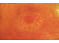
Q: I understand that the guidelines for following a patient on Plaquenil (hydroxychloroquine, Sanofi-Aventis) have changed. Can I still follow these patients in my office or do I need to comanage them with someone else?

A: “You can most definitely still follow them yourself, with the proviso that you are aware of the current best practices,” says Anthony Cavallerano, O.D., attending optometrist at VA Boston Health Care System. “The baseline testing and the ongoing monitoring of the patient can easily take place in an optometric office, especially because more and more optometric practices have spectral domain or other high-resolution OCT devices.”
Specifically, spectral domain OCT is one of three objective tests now recommended for routine screening of patients on Plaquenil. The other two are multifocal electroretinogram (mfERG) and fundus autofluorescence (FAF).
Why the change in the recommendations? Because “the risk of toxicity after years of [Plaquenil] use is higher than previously believed,” the study says.1 Also, better objective testing equipment has been developed since the previous guidelines were released in 2002, so it’s more important than ever to screen for early changes—and the earlier, the better.
Dr. Cavallerano says, “the new guidelines have thrown light on what we’ve all been doing for many years, namely subjective Amsler grid testing, fundus photography and sometimes fluorescein angiography, which may not hold up given the newer technologies.”
Although the new guidelines expose the limitations of Amsler grid testing, the recommendation for automated visual fields is intact. The guidelines mention 10-2 automated perimetry in particular. “When fields are performed independently, even the most subtle 10-2 field changes should be taken seriously and are an indication for evaluation by objective testing,” the new recommendations say.1
Regarding those objective tests, mfERG requires a special instrument that most optometrists and ophthalmologists don’t have, Dr. Cavallerano says.

New guidelines aim to prevent patients from ever developing bull’s-eye maculopathy.
However, many primary eye care doctors do have (or have access to) spectral domain OCT. (Time-domain OCT won’t serve the purpose, though. “Time domain OCT is just not sensitive enough to detect early changes in the retinal pigment epithelium and in the outer segments of the photoreceptors.”)
The other recommended objective test, fundus autofluorescence, can also be readily performed by optometrists. Many imaging systems, fundus cameras and SD-OCTs now have this capability using certain filters, Dr. Cavallerano says. “Any changes—even subtle defects—will be more apparent with fundus autofluorescence than with most conventional methods of imaging,” he says. “If an optometrist is going to do any of these tests, autofluorescence is the way to go.”
What early changes do you look for?
Using SD-OCT, “look for thinning in the outer retinal layers,” he says. “The loss of the inner and outer segment—that so-called inner-outer segment line—shows up fairly nicely in the parafoveal area.”
Using FAF, “look for increased autofluorescence of debris or detritus at the outer segments of the photoreceptors, which indicate areas of photoreceptor damage.”
If you do detect any of these early changes, “get the patient a retina consult as a matter of course,” Dr. Cavallerano says. “Then by all means communicate with the rheumatologist or internal medicine provider.”
The risk for Plaquenil toxicity increases after five years, and retinal changes take time to develop. So keep in mind that patients who have been switched to a newer medication could still demonstrate toxicity if they were once on Plaquenil.
1. Marmor MF, Kellner U, Lai TY, et al. American Academy of Ophthalmology. Revised recommendations on screening for chloroquine and hydroxychloroquine retinopathy. Ophthalmology. 2011 Feb;118(2):415-22.

