 |
History
A 71-year-old black male presented to the office for ongoing care following cataract surgery with anterior chamber lens implantation O.S. His chief complaint was poor vision and pain in the operated eye. His ocular history was unremarkable, and his systemic history was noncontributory. He reported compliance with his topical medications (ciprofloxacin, ketorolac and prednisolone acetate) and denied taking any new prescribed oral medication.
Diagnostic Data
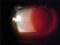
His best-corrected acuity was 20/400 O.D., light perception O.S. at distance and near. External examination was normal. The pupil could not be visualized in the left eye, but a reverse afferent defect was detectable. The pertinent anterior segment findings in the left eye are demonstrated in the photograph. There was a dense, hypermature cataract in the right eye.
Goldmann intraocular pressures measured 14mm Hg in both eyes. Peripheral and posterior pole dilated fundoscopy was within normal limits in the right eye. There was no view in the left.
 |
| An elderly black male presented with this anterior segment appearance O.S. |
Discussion
Additional testing included Seidels test to rule out a wound leak. Also, careful observation using the biomicroscope was needed to rule out hypopyon and postoperative endophthalmitis. B-scan was not considered at this time because of the concern that the newly placed IOL could be destabilized.
The diagnosis in this case was uveitis-glaucoma-hyphema (UGH) syndrome. This syndrome is characterized by inflammation after anterior chamber IOL implantation, which is classically caused by the haptics of the IOL itself. Misplaced or misdirected haptics from the anterior chamber IOL erode the tissues of the angle, causing bleeding and inflammation. The severity and intensity of the breakdown of the blood-aqueous barrier determine the extent of the clinical signs that a patient may demonstrate. These signs range from subclinical or minimally symptomatic inflammation to severe uveitis with glaucoma and hyphema (UGH syndrome).
A high incidence of this condition, associated with poor quality iris-supported and anterior chamber IOLs, occurred in the late 1970s and early 1980s. A much lower incidence of UGH syndrome exists today due in part to improved IOL technology and vastly decreased incidence of iris-supported and anterior chamber IOL use.14
The treatment for UGH is straightforward.2 Ask the patient to maintain bed rest with elevated head position to encourage hyphema settling. Antiplatelet analgesics or other medications known to increase the risk of bleeding should be avoided. Pain can be managed with acetaminophen or acetaminophen with codeine, and cycloplegia obtained with atropine 1% b.i.d. Address the inflammation with aggressive administration of topical prednisolone acetate 1% q2h. Lastly, reduce increased IOP via topical, oral, or combinations of topical and oral ocular hypotensive preparations (e.g., timolol, levobunolol, brimonidine, iopidine, acetazolamide).1,2 Avoid the prostaglandin class of agents (latanoprost, bimatoprost, travaprost, unoprostone) as they may exacerbate the inflammatory process.
Ultimately, the lens may have to be repositioned or removed.
Since a number of infections produce postoperative inflammation in IOL implantation, endophthalmitis must be ruled out. This infectious/inflammatory reaction begins within the first few postoperative days, and it typically includes pain, chemosis, hypopyon and corneal decompensation. Treatment of acute postoperative infectious endophthalmitis is governed by the National Eye Institutes Endophthalmitis Vitrectomy Study (EVS).5
1. Isaacs RT, Apple DJ. Evolution and pathology of intraocular lens implantation. In: Yanoff M, Duker JS. Ophthalmology. Philadelphia: Mosby, 1999:4.13.1-12.
2. Spaeth GL, Katz LJ, Moster MR, et al. Glaucoma: Postoperative glaucoma. In: Rhee DJ, Pyfer MF. The Wills Eye Manual. Philadelphia: Lippincott, Williams & Wilkins, 1999:263-6.
3. Cates CA, Newman DK. Transient monocular visual loss due to uveitis-glaucoma-hyphema (UGH) syndrome. J Neurol Neurosurg Psychiatry 1998 Jul;65(1):131-2.
4. Mamalis, N. Explanation of intraocular lenses. Curr Opin Ophthalmol 2000 Aug;11(4):289-95.
5. Wisniewski SR, Capone A, Kelsey SF, et al. Characteristics after cataract extraction or secondary lens implantation among patients screened for the Endophthalmitis Vitrectomy Study. Ophthalmology 2000 Jul;107(7):1274-82.
Vol. No: 143:01Issue:
1/15/2006

