 |
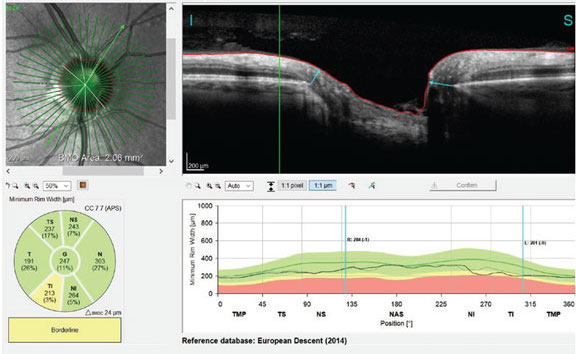
| Correctly locating Bruch's membrane opening is crucial in patients with high myopia. Photo: James Fanelli, OD. Click image to enlarge. |
Diagnosing glaucoma in highly myopic eyes can be challenging due to similar morphologic changes caused by the two diseases, including disc tilt in the optic nerve and enlarged peripapillary area. Examining the optic nerve in these patients through optical coherence tomography (OCT) may decrease diagnostic accuracy if scan results are not carefully and properly interpreted while considering the myopia status. Specifically, a recent study found that correct localization of Bruch’s membrane opening (BMO) is crucial in highly myopic patients, as this anatomical structure is used to define the optic nerve head (ONH) center and collect accurate RNFL thickness measurements.
Participants were gathered from a US study and a Korean clinic population, totaling 438 non-myopic healthy eyes from 263 subjects and 113 highly myopic eyes from 81 subjects. Of the total eyes, 397 had glaucoma (80 of the highly myopic eyes) and 154 did not (33 of the highly myopic eyes). High myopia was characterized as an axial length (AL) <26mm. All participants completed full ophthalmic examinations, including visual fields, coherence interferometry AL measurements, simultaneous stereophotography of the optic disc and spectral domain OCT of the macula. OCT obtained 24 radial scans and three circle scans centered on the ONH from each participant.
The data showed that the following factors increased the likelihood of BMO location inaccuracy: axial high myopia, larger AL, larger BMO tilt angle, a more oval BMO and glaucoma.
“In healthy eyes with and without axial high myopia, 9.1% and 1.7% had indiscernible BMOs while 54.5% and 87.6% were accurately segmented, respectively,” the researchers wrote in their study. They added that “36.4% and 10.7% of eyes with indiscernible BMOs were manually correctable. In glaucoma eyes with and without high myopia, 15% and 3.2% had indiscernible BMOs, 55% and 38.2% were manually corrected and 30% and 58.7% were accurately segmented without need for manual correction, respectively.”
The researchers concluded that BMO location inaccuracy was 2.4-times more likely in highly myopic eyes regardless of diagnosis. When monitoring or examining for signs of glaucoma in high myopes, OCT images must be carefully reviewed, and BMO location should be corrected, if possible, prior to using scan results to inform clinical decisions.
Rezapour J, Proudfoot JA, Bowd C, et al. Bruch’s membrane opening detection accuracy in healthy and glaucoma eyes with and without axial high myopia in an American and Korean cohort. Am J Ophthalmol. November 29, 2021. [Epub ahead of print]. |

