 |
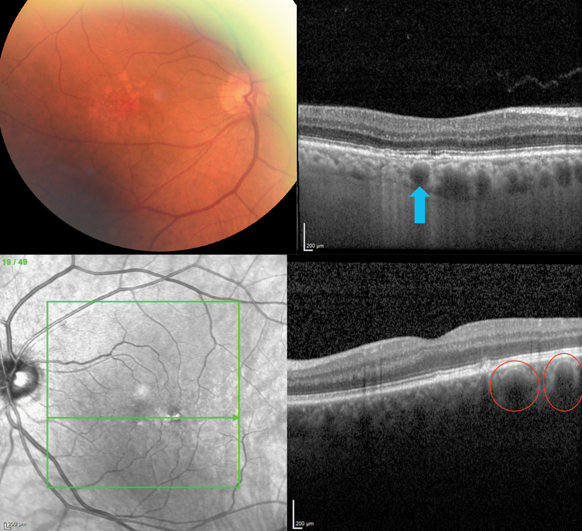
Choroidal vascularity index could have prognostic utility in AMD. Photo: Jessica Haynes, OD. Click photo to enlarge. |
Many studies have evaluated choroidal thickness in AMD with somewhat inconsistent results. The presence of subretinal drusenoid deposits (SDD) could be a factor, and in this study, researchers aimed to analyze the role of the choroid in early age-related macular degeneration (AMD) by analyzing the choroidal vascularity index in pure cohorts of patients with SDD or conventional drusen.
Sixty-nine eyes of 69 participants were included: 23 eyes with SDD, 22 eyes with conventional drusen and 24 controls. Comprehensive ophthalmologic examination and multimodal imaging including fundus photography, autofluorescence, near infrared reflectance and spectral domain optical coherence tomography (SD-OCT) were performed. Choroidal vascularity index processing was performed on a foveal horizontal SD-OCT scan with binarization and calculated as the ratio between luminal area and total area.
The authors did not find a difference between the total area between the three groups, but they found a significant difference in the luminal area of all three. They also found a significant reduction of the choroidal vascularity index of the SDD vs. the conventional drusen group, as well as reduced choroidal vascularity index of the conventional drusen group vs. controls.
“Based on histological studies on AMD eyes, primary damage is hypothesized to initiate at the choroidal level. Thus, it is reasonable to assume that the choroidal vascularity index is reduced in both the conventional drusen and SDD groups with respect to controls with a more pronounced reduction in eyes with SDD,” the authors explained.
Previous studies concluded that the luminal area was progressively more impaired in increasing severity from healthy controls to eyes with drusen, to eyes with SDD and to eyes with geographic atrophy. This study found similar results—a lower choroidal vascularity index in the conventional drusen group with respect to controls, and a lower choroidal vascularity index in the SDD group with respect to the conventional drusen group indicating that the choroidal vascularity index can be correlated to worsening AMD stages linked to phenotype, indicating progression risk. Thus, choroidal vascularity index could have prognostic implications in AMD, the authors suggested.
“In conclusion, we found that, in early AMD, the choroidal vascularity index in patients with pure phenotypes of SDD or conventional drusen was reduced with respect to controls, and the choroidal vascularity index was significantly reduced in SDD with respect to conventional drusen eyes,” the authors explained. “Our results were comparable with the studies with recruitment criteria similar to our investigation that eyes with SDD have more pronounced choroidal vascular alterations. SDD is a biomarker of AMD progression, and this work suggests that the mechanism for this is via reduction of the choroidal vascularity index.”
Abdolrahimzadeh S, Di Pippo M, Sordi E, et al. Subretinal drusenoid deposits as a biomarker of age-related macular degeneration progression via reduction of the choroidal vascularity index. Eye (Lond). June 23, 2022. [Epub ahead of print]. |

