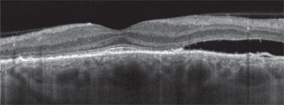
 A 52-year-old white female presented with a complaint of a sudden blind spot for the past few days. Upon examination, her left retina showed an elevated lesion near the macula, which looked like central serous retinopathy. But this semi-retired older woman is certainly not the usual high-stress, Type A young male—her only other worry is tennis elbow. What am I missing?
There’s been a longstanding association between central serous retinopathy (CSR), Type A personality and stress.1
“But if you ask such a patient, ‘Are you under any stress?’ that’s a loaded question,” says Jeffry Gerson, OD, of Olathe, Kan., who lectures frequently on retinal disorders. “This patient had sudden vision loss and she doesn’t know why. Of course she’s going to say she’s stressed.”
He adds, “And, even without that, how many people do you meet who say they don’t have any stress in their lives?”
Dr. Gerson says that the research on Type A personality, stress and CSR has never been conclusive, but there’s something else you should ask about in the history instead: systemic steroid use.
“Although central serous is idiopathic most of the time, steroid use is a strong risk factor,” he says. Research has shown that patients with CSR are three to 10 times more likely than control subjects to be using steroids.2
Be sure to ask about any kind of steroid use, not just oral steroids. “Ask about inhaled steroids for asthma, steroid injections for arthritis or a topical steroid ointment for a skin rash,” Dr. Gerson says.

| |
|
If a patient appears to have central serous retinopathy, ask about any type of steroid use. Also, Be sure to confirm the diagnosis with OCT or fluorescein angiography.
|
You may be surprised what you find. “I had a patient who had a hemorrhoid and started using steroid cream. Three or four days later, he developed blurred vision and had central serous,” he says. “It taught me a lesson—sometimes you have to ask very general questions in order to get a specific answer.”
After a detailed history, the next step is to clinically confirm the diagnosis of CSR with either optical coherence tomography (OCT) or fluorescein angiography (FA). CSR appears on OCT as a serous detachment or “blister” of subretinal fluid, and as a single “smoke stack” leakage on FA.
“The reason for performing OCT or FA is that a small choroidal neovascular membrane may mimic early central serous on clinical examination,” Dr. Gerson says. “Central serous is likely to improve and completely resolve after several weeks, whereas a choroidal neovascular membrane is likely to get worse with accompanying hemorrhage and exudates, which will cause significant vision loss.”
Next, if the patient with confirmed CSR is using or has recently used a systemic steroid, “it’s really important to inform the patient, as well as notify the primary care physician or specialist who prescribed the steroid—their rheumatologist managing the patient’s arthritis, for example—to educate them about the link between steroids and central serous.” Discuss the possibility of suspending or altering steroid therapy in the short term.
In the meantime, provide the patient with an Amsler grid to self-monitor her vision, and see her back in a month, Dr. Gerson says. (Tell her to come in immediately if she notices any worsening of vision.) The CSR will likely resolve on its own in a month or two.
1. Yannuzzi LA. Type-A behavior and central serous chorioretinopathy. Retina. 1987 Summer;7(2):111-31.
2. Nicholson B, Noble J, Forooghian F, Meyerle C. Central serous chorioretinopathy: update on pathophysiology and treatment. Surv Ophthalmol. 2013 Mar-Apr;58(2):103-26.

