|
A: Classified as the only member of the Stenotrophomonas genus, the bacterium Stenotrophomonas maltophilia is found in most soil and water environments, but it has only recently gained the attention of the eye care community as an emerging pathogen in corneal infection. This is possibly because clinicians are more thorough when patients present with a corneal ulcer and labs are going that bit further to identify and speciate cultures from patients, suggests microbiologist Simon Kilvington, Ph.D., of the University of Leicester, U.K.1
According to Clarke D. Newman, O.D., an officer of the AAO Section on Cornea, Contact Lenses and Refractive Technologies, there are several reasons for keeping a close watch on this bacterium.
“First, the incidence of this pathogen as a cause of ocular disease is increasing (tripling in incidence from 1970 to 1995) and is often nosocomial.” In fact, it entered the top 10 most common pathogens encountered in contact lens wear in Texas during that time period, Dr. Newman says.2
“Second, this pathogen can be found in isolate in the cases of contact lens patients. Third, it is often resistant to therapy, showing high MIC90 concentrations for most antibacterials.
Finally, it is a primary nutrient source for Acanthamoeba. So, when S. maltophilia is found, amoebic organisms could be lurking,” he says.3-5

|
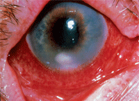
| Inferior corneal infiltrate in patient wearing contact lenses with bacterial keratitis.
|
In a recent ARVO presentation, Stenotrophomonas has been found as common lens case contaminant.7 Several clinical scenarios can be expected: “The sheer burden of organisms can initiate an immunological response in the cornea (directly or through endotoxin) leading to infiltrates,” Dr. Kilvington warns. “The presence of the bacterium in a storage case could allow the amoeba (introduced from the environment) to feed, replicate, adhere to the contact lens and become inoculated on to the cornea causing Acanthamoeba keratitis.”
So how do our patients acquire this bug? “The mode of acquisition is usually inadvertent contact with a contaminated source, which in most cases is some type of water supply,” says J.
James Thimons, O.D., of Fairfield, Conn. Dr. Thimons says that although it’s not a particularly aggressive organism, doctors must diagnose and kill it to prevent corneal infiltrative events.
“Treatment should be directed toward a typical gram-negative outcome,” he says. “Initial therapy is based on a fourth-generation fluoroquinolone. If that’s not satisfactory, I’ll go with fortified tobramycin while maintaining coverage with a fourth-generation fluoroquinolone.” Alternating the two drugs every half hour, Dr. Thimons expects the bug to stabilize within 24 hours and reverse within 48 hours.
Education on lens care, case replacement and avoidance of contaminated water is key in minimizing the risk for microbial events.
1. Denton M, Kerr KG. Microbiological and clinical aspects of infection associated with Stenotrophomonas maltophilia. Clin Microbiol Rev. 1998 Jan;11(1):57-80.
2. Penland, RL, Wilhemus KR. Stenotrophomonas maltophilia ocular infections. Arch Ophthalmol. 1996 Apr;114(4):433-6.
3. Bottone EJ, Madayag RM, Qureshi MN. Acanthamoeba keratitis: synergy between amebic and bacterial co-contaminants in contact lens care systems as a prelude to infection. J Clin Microbiol. 1992 Sep;30(9):2447-50.
4. Clark BJ, Harkins LS, Munro FA, Devonshire P. Microbial contamination of cases used for storing contact lenses. J Infect. 1994 May;28(3):293-304.
5. Donzis PB, Mondino BJ, Weissman BA, Bruckner DA. Microbial contamination of contact lens care systems. Am J Ophthalmol. 1987 Oct 15;104(4):325-33.
6. Nikolic M, Kilvington S, Cheung S, et al. Survival and Growth of Stenotrophomonas maltophilia in Multipurpose Contact Lens Solutions. Invest. Ophthalmol. Vis. Sci. 2010 51:1540.
7. Wiley L, McAllister M, Wiley LA, et al. Biofilm Bacterial Diversity: Association With Disease Severity in Contact Lens Related Keratitis. Poster D972. Presented at ARVO 2011.

