A 26-year-old female presented for an evaluation of blurry vision and floaters that began four weeks prior. She stated that after eating a ghost pepper, she developed an allergic reaction and became unconscious. She was taken to the hospital where she developed aspiration pneumonia, myocarditis and hypotension. She was subsequently intubated and given 1mL epinephrine injection for the evident type 1 hypersensitivity reaction. Since then, she reported, her blood pressure has returned to normal levels and her other systemic problems have resolved. She no longer notices floaters but still complains of blurry vision in her left eye.
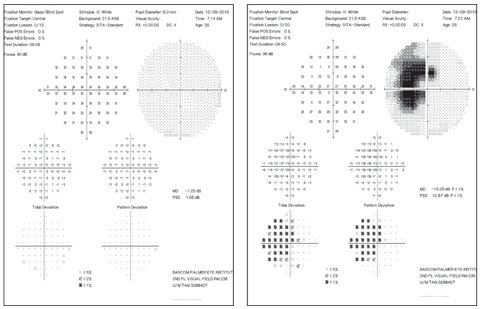
Upon examination, the patient’s best-corrected vision was 20/20 OD and 20/30 OS. Confrontational visual fields were full in the right eye and revealed a temporal paracentral scotoma in the left. Evidence of the temporal paracentral visual field scotoma was also found in Amsler grid and visual field testing. Her pupils were equally round and reactive to light; no afferent pupillary defect was found. Intraocular pressure (IOP) was 13mm Hg in both eyes. Slit lamp examination of both eyes was unremarkable.
 |
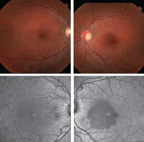
| Figs.1 and 2. At top, fundus photos of the right and left eye of our patient. Below are red-free versions. Look carefully at the left eye images. Click image to enlarge. |
A dilated fundus exam of the right eye was normal. The left eye revealed very subtle pigmentary changes nasal to the fovea (Figures 1 and 2). The remainder of the fundus exam was otherwise normal. Red-free fundus photography was performed (Figure 3) as well as SD-OCT (Figure 4). Multifocal ERGs (mfERG) and FAF was also obtained.
Take the Retina Quiz
1. What are the findings on the retinal photograph of the left eye?
a. Trace optic nerve pallor.
b. Choroidal neovascular membrane.
c. Macular edema.
d. Essentially normal.
2. What layer(s) of the retina seem to be affected on the OCT of the left eye?
a. Outer nuclear layer.
b. Outer plexiform layer.
c. Ellipsoid zone.
d. All of the above.
3. What is the correct diagnosis?
a. Branched artery occlusion.
b. Acute macular neuroretinopathy.
c. Pattern dystrophy.
d. Multiple evanescent white dot syndrome.
4. What is the appropriate treatment?
a. Observe.
b. Topical steroids.
c. Anti-VEGF injections.
d. Refer for an MRI.
5. What are known risk factors for this condition?
a. Sun exposure.
b. High cholesterol.
c. Sympathomimetics.
d. Genetic predisposition.
 |
| Fig. 3. Can this SD-OCT of the patient’s left eye help yield the diagnosis? Click image to enlarge. |
Diagnosis
Even though the fundus of the left eye appeared normal, subtle RPE changes could be seen. The OCT of the left eye showed obvious ellipsoid zone disruption as well as nasal hyper-reflective OPL/ONL and ONL thinning. The red-free photos of the left eye showed a dramatic paracentral dark grey, nasal hyporeflective lesion pointing toward the foveal center. The multifocal ERG was normal in the right eye, but indicated impaired responses in the retinal area around the blind spot in the left.
Based on the history, clinical findings and imaging studies, we suspected that our patient had acute macular neuroretinopathy (AMN), an idiopathic, rare disease that most commonly affects young to middle-aged females.1-3 It was initially believed to involve the inner retina, but since the advent of SD-OCT, it is now known to be located in the outer retina with characteristic ellipsoid zone disruption, which has forced authors to refer to AMN as acute macular outer retinopathy (AMOR).2,3,5
 |
| Fig. 4. What information can be gleaned from this patient’s visual fields? Click image to enlarge. |
Discussion
The condition can present unilaterally or bilaterally with acute onset of a paracentral scotoma and photopsias, typically without involvement of central visual acuity.1,3Paracentral location has been proposed simply due to the increased capillary network in this area.4 The fundus can appear normal as was the case with our patient, but days to months after initial onset of symptoms there are usually one or more red/brown perifoveal petalloid-like or wedge-shaped lesions with the tip pointing towards the fovea.1,3 Though these may be difficult to see on clinical exam, red-free light and near-infrared reflectance may allow better visualization.5 OCT of these lesions typically reveal disruptions of the ellipsoid zone.1
Many AMN cases have been associated with an acute viral illness preceding the onset of symptoms. It has also been reported to occur after the use of oral contraceptives, IV contrast medium injections, excessive amounts of caffeine intake, allergic reactions to prawns followed by administration of vasoconstricting drugs, shock, trauma, severe blood loss and episodes of acute systemic hypotension.2-4 Our patient noted her symptoms following an epinephrine injection.
The pathophysiology of AMN is not completely understood, but it may occur as a result of an acute episode of choroidal ischemia.2 However, many authors refute this notion and argue that it is due to an acute retinal ischemic event, and due to the fact that central vision is often unaffected, it is assumed that the ischemia is to the deep capillary plexus of the central retinal artery rather than to the choroid or choriocapillaris.3 On high magnification fluorescein angiography, dilation of the deep capillary plexus has been seen, which further supports an ischemic etiology.4 In addition, it is not uncommon to see the presence of one or more flame-shaped hemorrhages, which further supports a vascular occlusive event.2,4
Comorbidities
There may be an association of AMN with multiple evanescent white dot syndrome (MEWDS), acute idiopathic blind spot enlargement syndrome (AIBSE) and a condition called pseudo presumed ocular histoplasmosis (pseudoPOHS). Some authors recommend these disorders be grouped under the umbrella term of acute zonal occult outer retinopathy (AZOOR).3
Paracentral acute middle maculopathy (PAMM) is a newly identified variant of AMN that involves the middle layers of the retina, above and below the OPL.4 Type 1 AMN, now know as PAMM, refers to hyper-reflectivity of the OPL/INL on OCT with subsequent INL thinning.4,6 It is usually seen in older men with vascular disease around 60 years of age, but now is also being seen in young women.4,6 Type 2 AMN, which is more common in young, healthy women around 30 years of age, involves the OPL/ONL. When hyper-reflectivity on OCT of the OPL/ONL resolves, evidence of ONL and subsequent ellipsoid zone thinning is seen, because 10% to 15% of the oxygen supply to the photoreceptors is by the deep capillary plexus.4,5,6
AMN tends to be non-recurrent and self-resolving, but symptoms may persist. There is no current treatment for this condition.1,3
We chose to closely monitor our patient. She was seen monthly for three months and showed steady improvement back to 20/20. However, a paracentral scotoma persisted on Amsler grid. Interestingly, the pigmentary changes became more evident.
Dr. Brafman is an optometric resident at the Bascom Palmer Eye Institute in Miami.
|
1. Kirkpatrick C. Acute Macular Neuroretinopathy (AMN). University of Iowa Health Care: Ophthalmology and Visual Sciences (2015). http://www.eyerounds.org/atlas/pages/Acute-macular-neuroretinopathy/index.htm. Accessed April 7, 2016. 2. Hughes E, Siow Y, Hunyor A. Acute macular neuroretinopathy: anatomic localization of the lesion with high-resolution OCT. Eye. 2009;23:2132-4. 3. Douglas I, Cockburn D. Acute macular neuroretinopathy. Clinical and Experimental Optometry. 2003;2:121-6. 4. Sarraf D, Rahimy E, Fawzi A, et. al. Paracentral acute middle maculopathy: a new variant of acute macular neuroretinopathy associated with retinal capillary ischemia. Jama Ophthalmol. 2013;131(10):1275-87. 5. Joseph A, Rahimy E, Freund B, Sarraf D. Multimodal Imaging in Acute Macular Neuroretinopathy. Retinal Physician 2013 June;10:40-4. 6. Tsui, I, Sarraf D. Paracentral Acute Middle Maculopathy and Acute Macular Neuroretinopathy. Ophthalmic Surg Lasers Imagine Retina. 2013;44(6):S33-S35. |
Retina Quiz Answers:
1) d; 2) d; 3) b; 4) a; 5) c.

