When a contact lens wearer presents with a painful red eye and a suspicious corneal infiltrate that is gray and “wispy,” are there any clinical indicators that help initially differentiate between bacterial and fungal (non-bacterial) etiologies?

If left untreated, a corneal infiltrate could result in a host of morbidities, which is why a prompt diagnosis and proper treatment plan are imperative. While many factors must be considered when examining an eye with a corneal ulceration, a definitive diagnosis can only be made with the help of a culture.
A study presented at the 2010 ARVO Annual Meeting confirmed this—participating corneal specialists were able to correctly differentiate between bacterial and fungal presentations just 65.5% of the time.1 Doctors’ ability to recognize bacterial vs. fungal ulcers was put to the test using photographs of confirmed diagnoses. Although the participants used traits such as infiltrate border appearance, surrounding stromal haze and presence of hypopyon, gram stains, genus and species were accurately predicted only 48.2%, 25.7% and 20.9% of the time, respectively.
It’s clear—to avoid misdiagnosis of corneal ulcers, eye care practitioners should rely primarily on laboratory testing. But, James V. Aquavella, M.D., professor of ophthalmology at the
University of Rochester Flaum Eye Institute, considers history to be an important additional aspect in establishing a presumptive diagnosis.
“Contact lens wear or a history of trauma to the corneal epithelium both facilitate penetration of the organism across the epithelial barrier and may be helpful in elevating the index of suspicion,” he says. Other risk factors for fungal keratitis include:
• Diabetes and other immunosuppressive conditions.

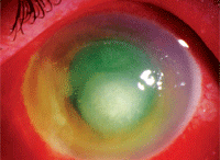
Central corneal ulcer with hypopyon in a patient with fungal infection.
• Travel to warm, humid climates.
• Contact with vegetable matter.
• Contact with farm animals.
• Long-term instillation of antibiotic and steroid drops.
• Failed antibiotic therapy.
Dr. Aquavella warns that the course of the infective process of fungal keratitis may be slower than that of acute bacterial ulceration; this may be attributed to initial misdiagnosis as bacterial and prolonged antibiotic therapy. In these instances, preliminary cultures may be negative or even positive for normal flora.
So, what symptoms warrant further testing?
“Pain, tearing, photophobia and significant bulbar conjunctiva injection are often associated with fungal ulcerations,” says Dr. Aquavella. “Severe anterior iritis with intense flare, leading to the development of a plasmoid aqueous or frank hypopyon level are also characteristic of fungal disease.”
Slit lamp findings of infiltrates with fungal etiologies reveal a stromal ulceration with elevated, raised or rolled edges vs. the sloping edges typically seen in bacterial lesions. Surrounding particulate matter in the stroma, feathery subepithelial extensions from the confines of the ulceration and frank satellite lesions are closely associated with fungal lesions, says Dr. Aquavella. When the cause of the ulcer is mold, the epithelium can be intact or ulcerated and non-suppurative stroma with feathery infiltrates can be noted. But, yeast causes ulcerations and suppuration in the stroma (focal or diffuse).
“In some cases, an immune ring may be present, necessitating a differential diagnosis with the less frequent but equally devastating Acanthamoeba keratitis.” When cultures are deemed appropriate, corneal scraping should be plated directly onto chocolate, blood and Sabouraud’s media (non-medicated). This will identify most bacteria and fungi.
1. Dalmon CA, Mascarenhas JM, Lalitha P, et al. Clinical Differentiation of Bacterial and Fungal Corneal Ulcers. Presented at ARVO 2010. Abstract: 2880/D1104.

